Archives

Play Video

Watch how blood flows through the heart

(Credit: Getty Images)

A new simulation shows how blood flows through the heart.

This view inside a beating heart is one not seen—or even seeable—until now, according to Tayfun Tezduyar.

Tezduyar, professor of mechanical engineering at Rice University’s George R. Brown School of Engineering, says such visualizations can help clinicians understand the mechanisms that push blood through the body and fix things when they go awry.

Tezduyar is an expert in modeling interactions between fluids and structures whose work encompasses biomechanics, including the circulatory system, and large-scale systems like parachutes, wind turbines, and ground vehicles.

In this research, Tezduyar and his colleagues at Waseda University in Japan tackled what he says is one of the toughest problems in biomechanics: a way to accurately characterize the flow of blood around and through the heart’s valves, taking into account the flow details near the surfaces of the valve’s three leaflets along the way.

The animation shows two views of the ventricle-valve-aorta, top and side, and reveals in great detail how the ventricle fills with blood, how quickly the three-leaf valve opens to relieve the pressure, and the turbulent flow of blood as it pushes through.

“This is a computational technology that no one else in the world currently has with this level of accuracy near the leaflet surfaces,” Tezduyar says. “We’ve become aware recently that people at the Texas Medical Center are asking for CFD (computational fluid dynamics) simulations of the cardiovascular system. I think perhaps they meant what happens just in the aorta, and that would have been a lot easier. But we went a little beyond.”

Tezduyar says the simulation incorporates the valve mechanism and fluid flow through the aorta and its three branch arteries.

“That’s a very difficult computation,” he says. “When someone needs to make decisions based on a simulation, the main issue is how accurate and reliable it is. The opening and closing of the valves make it extremely complex, and generally the more complex a simulation becomes, the less reliable it could be.

“What we’ve done is keep both the complexity and accuracy,” he says. “It was painstaking work. Modeling three-leaflet valves that touch each other, fully close off blood flow and then open again, and doing that with accurate representation of the near-leaflet flow patterns, is a new and challenging class of problems in fluid mechanics.”

He says the model is detailed enough to capture the spiraling motion of blood in the aorta.

“We need to represent the flow patterns on these walls correctly to evaluate shear stress on the arterial walls, which is connected to the risk of aneurisms,” says Tezduyar, who co-leads the Team for Advanced Flow Simulation and Modeling with Kenji Takizawa, a professor in the School of Creative Science and Engineering at Waseda University.

“This is valuable information for doctors looking for ways to cure heart disease,” he says. “For instance, the model would help them evaluate replacement valves, including the geometry, thickness, and material.”

The simulation of a working ventricle-valve-aorta represents calculations first presented by Tezduyar and his Waseda colleagues in 2020.

Takizawa was a research associate at Rice beginning in 2007. His research associate, Takuya Terahara, who also spent time at Rice as a visiting PhD student in 2018, produced the animation. Tezduyar says he and Takizawa are amenable to discussing with doctors long-term collaborative work on patient-specific simulations.

Source: Rice University
Play Video

Custom 3D-printed heart valves fit perfectly

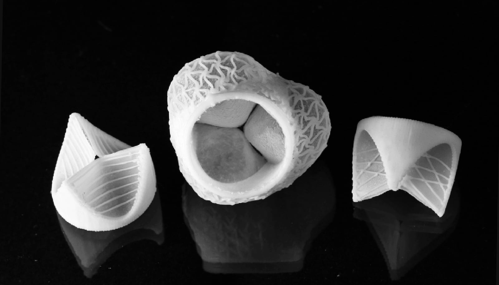
Patient-specific shaped heart valves. (Credit: Fergal Coulter/ETH Zurich)

Custom-made artificial heart valves made with 3D printing could help meet an aging population’s growing demand for replacement heart valves, according to new research.

The human heart has four chambers, each equipped with a valve to ensure blood flow in one direction only. If any of the heart valves are leaking, narrowed or distended (or even ruptured), the blood runs back into the atria or ventricles, putting the entire heart under severe strain. In the worst case, this can lead to arrhythmia or even heart failure.

Depending on the severity of the defect, artificial heart valves can remedy the problem. Over the next few decades, demand for this type of surgery is likely to soar in many parts of the world due to the aging population, lack of physical exercise, and poor diet. It is estimated that around 850,000 people will require artificial heart valves in 2050. Researchers have been looking for an alternative to the replacement heart valves currently in use.

Unlike conventional heart valves, the silicone heart valve can be tailored more precisely to the patient, as the researchers first determine the individual shape and size of the leaky heart valve using computer tomography or magnetic resonance imaging. This makes it possible to print a heart valve that fits the patient’s heart chamber perfectly.

The researchers use the images to create a digital model and a computer simulation to calculate in advance the forces acting on the implant and its potential deformation. The material used is also compatible with the human body, while the blood flow through the artificial heart valve is as good as with conventional replacement valves.

Heart surgeons have traditionally used implants that consist either of hard polymers or animal tissue (from cows or pigs) combined with metal frames. To prevent the body rejecting these implants, patients have to take life-long immunosuppressants or anticoagulants, which have significant undesirable side effects.

Easier, cheaper, better

In addition, conventional replacement valves have a very rigid geometric shape, making it challenging for surgeons to ensure a tight seal between the new valves and the cardiac tissue.

“The replacement valves currently used are circular, but do not exactly match the shape of the aorta, which is different for each patient,” says co-lead author Manuel Schaffner, a former doctoral student of André Studart, professor for complex materials at ETH Zurich. On top of that, manufacturing artificial heart valves is both expensive and time-consuming.

The new type of silicone heart valves gets around this problem. It only takes about an hour and a half for researchers to produce such a valve with a 3D printer. By contrast, it takes several working days to make an artificial heart valve by hand from bovine material. Production with 3D printers could also speed up: a battery of printers could, for example, produce dozens or even hundreds of valves every day.

Making new heart valves

First, the scientists create a negative impression of the valve. They spray silicone ink onto this impression in the shape of a three-pointed crown, which forms the valve’s thin flaps.

In the next step, an extrusion printer deposits tough silicone paste to print specific patterns of thin threads on their surface. These correspond to collagen fibers that pass through natural heart valves. The silicone threads reinforce the valve flap and extend the life of the replacement valve. The researcher print the root of the blood vessel connected to the heart valve using the same procedure and at the end they cover it with a net-shaped stent, which is necessary for connecting the silicone valve replacement to the patient’s cardiovascular system.

Initial tests have produced very promising results for the new valve’s function. The material scientists’ goal is to extend the life of these replacement valves to 10-15 years. This is how long current models last in patients before they need to be exchanged.

“It would be marvelous if we could one day produce heart valves that last an entire lifetime and possibly even grow along with the patient, so that they could also be implanted in young people as well,” says Schaffner.

However, it will still take at least 10 years before the new artificial heart valves come into clinical use, as they first have to go through exhaustive clinical trials.

Co-lead author Fergal Coulter, a postdoc in Studart’s group who developed the 3D printers that produce the heart valves, is currently working on the further development of the silicone heart valve. “These experiments are needed to make sure that the technology has any chance at all of being used in human patients,” he stresses.

Coulter is also continuing to experiment with new materials that could extend the life of the heart valves.

However, either an industrial partner or possibly a spin-off is needed to make the heart valve commercially available on the market. “As a research group, we are unfortunately unable to provide a seamless offering from the first experiment to the first application in the human body,” Schaffner emphasizes.

The research appears in the journal Matter. Additional researchers from ETH Zurich and the South African company SAT contributed to the work.

Source: ETH Zurich

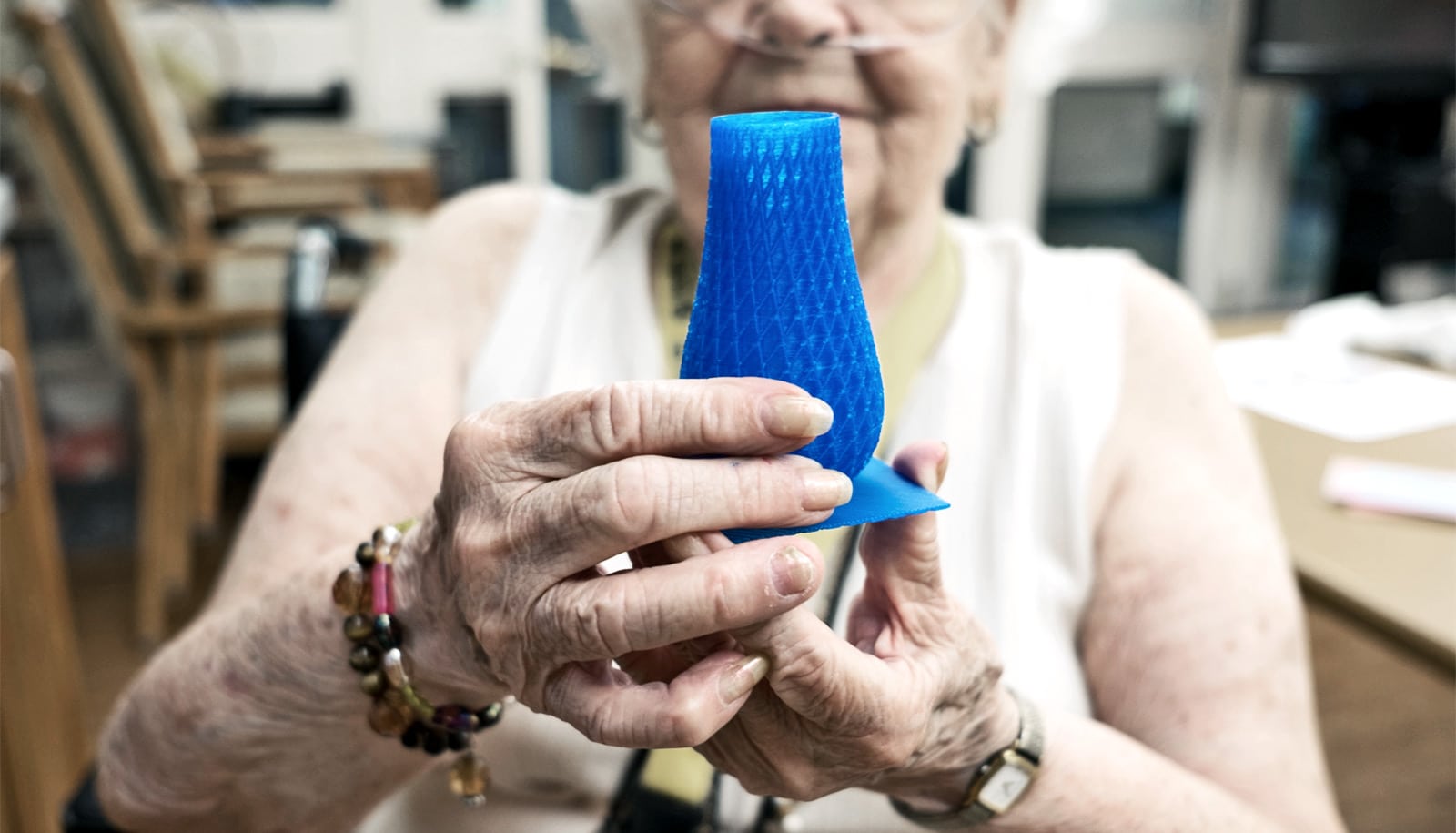
  • 3D printing lets long-term care residents get creative