Archives

Play Video

Genome ‘atlas’ pinpoints driver mutations that cancers share

(Credit: Getty Images)

Researchers have completed the most comprehensive study of whole cancer genomes to date.

The work significantly improves our fundamental understanding of cancer and signposts new directions for its diagnosis and treatment, the researchers report.

For the new analysis in the journal Nature, more than 700 researchers examined more than 2,600 samples from 38 cancer types. The cancers range from common ones like colorectal and breast cancers to rare cancer types including pancreatic and brain cancers.

This analysis has enabled researchers to create the first complete atlas of genomes to compare the cancers and find the common mutations between them.

The Pan Cancer Analysis of Whole Genomes or the Pan-Cancer Project (PCAWG) created the atlas. PCAWG is a collaboration between groups from the International Cancer Genome Consortium who agreed to put thousands of sets of patient genome data together and reanalyze these samples using cloud computing.

The mutations have all been combined into a sort of catalogue. The catalogue, which is already available online, allows doctors and researchers from all over the world to look things up, consult with, and find information about the cancer of a given patient.

Mapping driver mutations

“Most previous major studies have focused on the protein coding 2% of the genome. We have studied and analyzed the whole genome, and our analyses of mutations that are affecting cancer genes have enabled us to genetically explain 95% of the cancer occurrences we have studied by means of mutations,” says coauthor Joachim Weischenfeldt, associate professor at the Biotech Research & Innovation Centre at the University of Copenhagen and the Finsen Laboratory at Rigshospitalet.

“So, if you know which mutations have caused cancer, the so-called driver mutations, you will be able to better tailor a treatment with the most suitable drugs or design new drugs against the cancer. Precision medicine is completely dependent on the mapping of driver mutations in each cancer, in relation to diagnosis, prognosis, and improved treatment,” says coauthor Jakob Skou Pedersen, a professor at the Bioinformatics Research Centre and in the clinical medicine department at Aarhus University and Aarhus University Hospital.

Researchers found on average any given tumor has four to five key mutations—changes to the genetic blueprint—that are responsible for driving that disease. Those mutations can vary a great deal for each cancer type.

Previously researchers were aware of one or two drivers. Researchers say understanding that number and realizing the complexity in each patient is an important step in working out where else to look when diagnosing cancer patients.

“It is quite surprising that almost all of them have the same number of driver mutations. However, it is consistent with theories that a cancerous tumor needs to change a certain number of mechanisms in the cell before things start to go wrong,” says Pedersen.

This atlas provides a solid foundation for understanding which genes and which pathways may be damaged in each cancer type, says Sean Grimmond, chair in cancer medicine at the University of Melbourne.

“This research will help identify what types of genetic test are needed for each cancer type—filling in potential existing gaps that we did not even know were there,” Grimmond says. “It demonstrates better than ever before how similar damage can cause cancer in different tissues—implications mean that for example a breast cancer drug could be effectively used to treat an esophageal cancer.”

The atlas also identifies patterns of damage across various cancer types, which provides insight for challenging cancers where the tissue of origin is unknown.

“If we don’t understand where a cancer comes from, we can’t even rely on traditional clinical approaches to treatment,” Grimmond says.

Untangling the cancer genome for better treatment

Having a harmonized dataset enables international researchers to learn from one cancer treatment and applies those findings to another using a cloud computing portal.

The researchers say further research with much larger datasets is required to move precision medicine toward becoming a reality.

“This work is helping to answer a long-standing medical difficulty, why two patients with what appear to be the same cancer can have very different outcomes to the same drug treatment,” says Peter Campbell, member of the Pan-Cancer Project steering committee and head of cancer, aging, and somatic mutation at the Wellcome Sanger Institute in the UK.

“We show that the reasons for these different behaviors are written in the DNA. The genome of each patient’s cancer is unique, but there are a finite set of recurring patterns, so with large enough studies we can identify all these patterns to optimize diagnosis and treatment,” Campbell says.

“Cancer is a genetic disease, and the type of mutations is often more important than where the cancer originates in the body. This means that we need to think of cancer not just as a tissue-specific disease, but rather look at it based on genetics and the mutations it has,” says Weischenfeldt.

“For example, we may have a type of breast cancer and prostate cancer where the driver mutations are similar. This means that the patient with prostate cancer may benefit from the same treatment as the one you would give the breast cancer patient, because the two types share an important driver mutation.”


Source: University of Melbourne, University of Copenhagen

  • 1 number could make the hunt for cancer cells easier
  • Play Video

    A.I. boosts breast cancer detection accuracy

    (Credit: Getty Images)

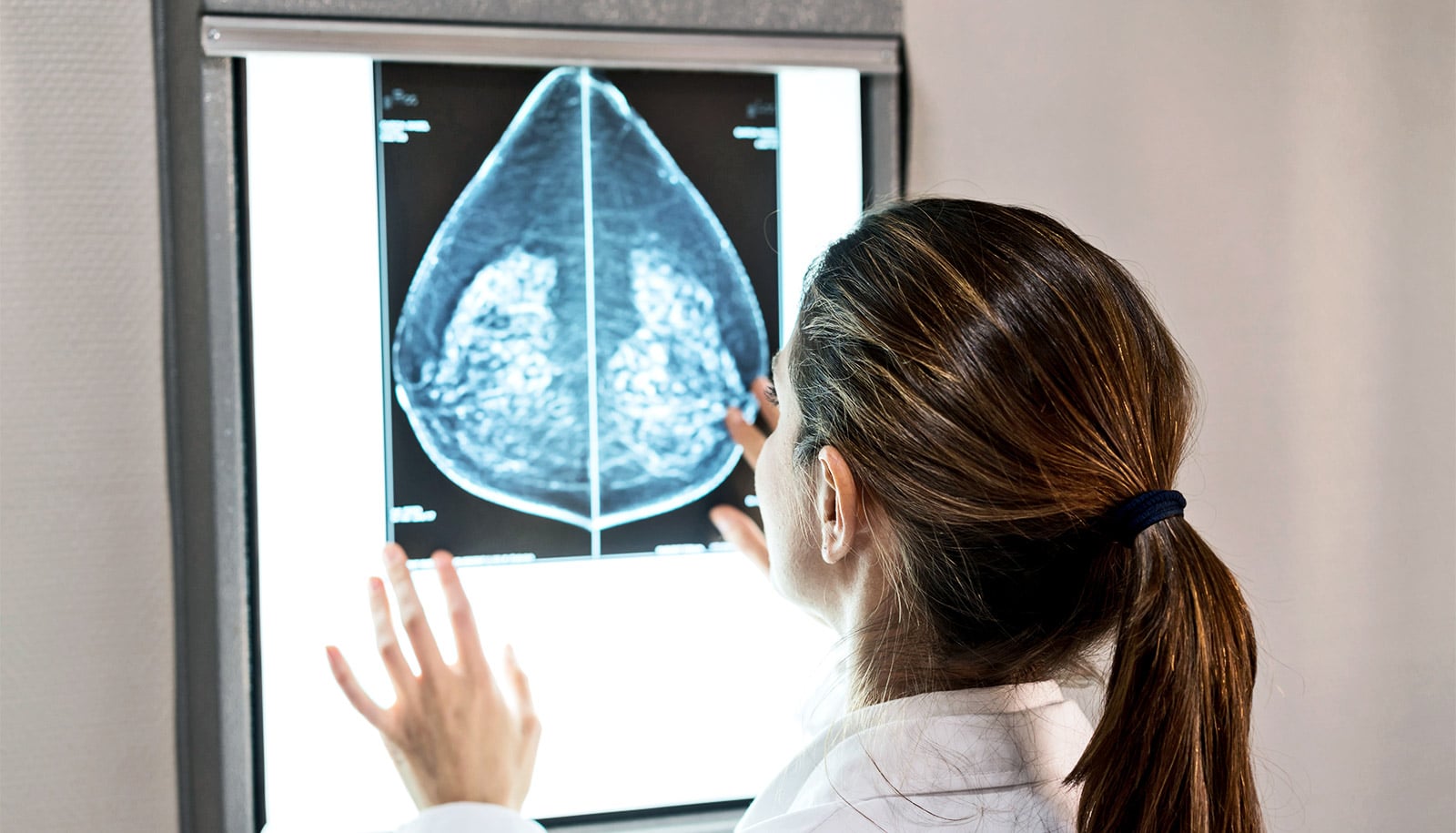
    An artificial intelligence tool—trained on roughly a million screening mammography images—can identify breast cancer with approximately 90% accuracy when combined with radiologist analysis, a new study finds.

    The study examined the ability of a type of artificial intelligence (AI), a machine learning computer program, to add value to the diagnoses a group of 14 radiologists reached as they reviewed 720 mammogram images.

    “The ultimate goal of our work is to augment, not replace, human radiologists.”

    “Our study found that AI identified cancer-related patterns in the data that radiologists could not, and vice versa,” says senior study author Krzysztof Geras, assistant professor in the radiology department at New York University’s Grossman School of Medicine.

    “AI detected pixel-level changes in tissue invisible to the human eye, while humans used forms of reasoning not available to AI,” adds Geras, also an affiliated faculty member at the Center for Data Science. “The ultimate goal of our work is to augment, not replace, human radiologists.”

    In 2014, women (without symptoms) in the United States got more than 39 million mammography exams to screen for breast cancer and determine the need for closer follow-up. Women whose test results yield abnormal mammography findings are referred for biopsy, a procedure that removes a small sample of breast tissue for laboratory testing.

    Three images of breast tissue side by side. The first is black and white, the second has spots of green, the third has spots of red.
    The AI tool learned to predict which lesions were likely malignant (red heat map) or likely benign (green heat map), with potential to aid radiologists in the diagnosis of breast cancer. (Credit: NYU School of Medicine)

    In the new study, the research team designed statistical techniques that let their program “learn” how to get better at a task without being told exactly how. Such programs build mathematical models that enable decision-making based on data examples fed into them, with the program getting “smarter” as it reviews more and more data.

    Modern AI approaches, which take inspiration from the human brain, use complex circuits to process information in layers, with each step feeding information into the next, and assigning more or less importance to each piece of information along the way.

    The authors of the current study trained their AI tool on many images matched with the results of biopsies performed in the past. Their goal was to enable the tool to help radiologists reduce the number of biopsies needed moving forward. This can only be achieved, says Geras, by increasing the confidence that physicians have in the accuracy of assessments made for screening exams (for example, reducing false-positive and false-negative results).

    For the current study, the research team analyzed images collected as part of routine clinical care over seven years, sifting through the collected data and connecting the images with biopsy results. This effort created an extraordinarily large dataset for their AI tool to train on, the authors say, consisting of 229,426 digital screening mammography exams and 1,001,093 images. Most databases the researchers used in studies to date have been limited to 10,000 images or fewer.

    Thus, the researchers trained their neural network by programming it to analyze images from the database for which cancer diagnoses had already been determined. This meant that researchers knew the “truth” for each mammography image (cancer or not) as they tested the tool’s accuracy, while the tool had to guess. The researchers measured accuracy in the frequency of correct predictions.

    In addition, the researchers designed the study AI model to first consider very small patches of the full resolution image separately to create a heat map, a statistical picture of disease likelihood. Then the program considers the entire breast for structural features linked to cancer, paying closer attention to the areas flagged in the pixel-level heat map.

    Rather than have the researchers identify image features for their AI to search for, the tool is discovering on its own which image features increase prediction accuracy. Moving forward, the team plans to further increase this accuracy by training the AI program on more data, perhaps even identifying changes in breast tissue that are not yet cancerous but have the potential to be.

    “The transition to AI support in diagnostic radiology should proceed like the adoption of self-driving cars—slowly and carefully, building trust, and improving systems along the way with a focus on safety,” says first author Nan Wu, a doctoral candidate at the Center for Data Science.

    The study appears in IEEE Transactions on Medical Imaging.

    Additional coauthors are from NYU, SUNY Downstate College of Medicine, the University of Cambridge, and Jagiellonian University.

    Support for the work came, in part, from the National Institutes of Health. The model used in this study has been made available to the field to drive innovation.

    Source: New York University