Permanent changes in bones result from giving birth and breastfeeding, according to an analysis of primates.
“Our findings provide additional evidence of the profound impact that reproduction has on the female organism, further demonstrating that the skeleton is not a static organ, but a dynamic one that changes with life events,” explains Paola Cerrito, who led the research as a doctoral student in New York University’s department of anthropology and College of Dentistry.
Specifically, the researchers found that calcium, magnesium, and phosphorus concentrations are lower in females who have experienced reproduction. These changes are linked to giving birth itself and to lactation.
However, they caution that while other clinical studies show calcium and phosphorus are necessary for optimal bone strength, the new findings do not address overall health implications for either primates or humans. Rather, they say, the work illuminates the dynamic nature of our bones.
“A bone is not a static and dead portion of the skeleton,” notes anthropologist Shara Bailey, an author of the study in PLOS ONE. “It continuously adjusts and responds to physiological processes.”
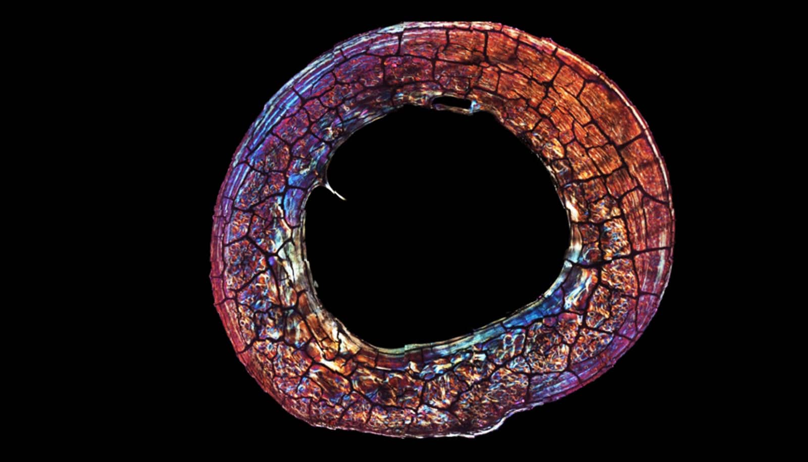
It’s been long-established that menopause can have an effect on bones. Less clear is how preceding life-cycle events, such as reproduction, can influence skeletal composition. To address this, the researchers studied the primary lamellar bone—the main type of bone in a mature skeleton. This aspect of the skeleton is an ideal part of the body to examine because it changes over time and leaves biological markers of these changes, allowing scientists to monitor alterations during the life span.
In the study, the researchers examined the growth rate of lamellar bone in the femora, or thigh bones, of both female and male primates who had lived at the Sabana Seca Field Station in Puerto Rico and died of natural causes. Veterinarians at the field station had monitored and recorded information on these primates’ health and reproductive history, allowing the researchers to match bone-composition changes to life events with notable precision.
Cerrito and her colleagues used electron microscopy and energy-dispersive X-ray analysis—common ways to gauge the chemical composition of tissue samples—to calculate changes in concentrations of calcium, phosphorus, oxygen, magnesium, and sodium in the primates’ bones.
Their results show different concentrations of some of these elements in females who gave birth compared males as well as females who did not give birth. Specifically, in females who gave birth, calcium and phosphorus were lower in bone formed during reproductive events. Moreover, there was a significant decline in magnesium concentration during these primates’ breastfeeding of infants.
“Our research shows that even before the cessation of fertility the skeleton responds dynamically to changes in reproductive status,” says Cerrito, now a research fellow at ETH Zurich. “Moreover, these findings reaffirm the significant impact giving birth has on a female organism—quite simply, evidence of reproduction is ‘written in the bones’ for life.”
Coauthors of the study are from NYU, Texas State University, and Brown University.
The research had support from the National Science Foundation and the National Institutes of Health.
Source: NYU