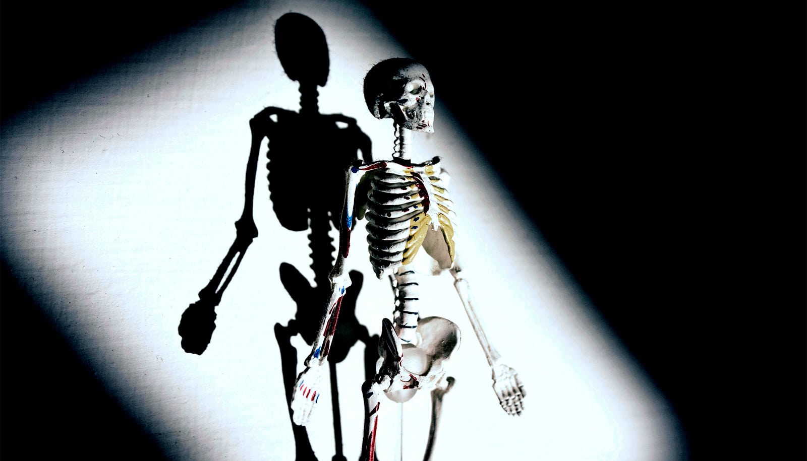
Researchers have developed ultra-thin wireless devices called osseosurface electronics that grow to the surface of bone and could someday help monitor bone health and healing over long periods.
“As a surgeon, I am most excited about using measurements collected with osseosurface electronics to someday provide my patients with individualized orthopedic care—with the goal of accelerating rehabilitation and maximizing function after traumatic injuries,” says David Margolis, an assistant professor of orthopedic surgery in the University of Arizona College of Medicine-Tucson and orthopedic surgeon at Banner University Medical Center Tucson.
Margolis is co-senior author of a paper describing the devices in Nature Communications.

Fragility fractures associated with conditions like osteoporosis account for more days spent in the hospital than heart attacks, breast cancer, or prostate cancer.
Although not yet tested or approved for use in humans, the wireless bone devices could one day be used not only to monitor health, but to improve it, says co-senior author Philipp Gutruf, an assistant professor of biomedical engineering and faculty fellow in the College of Engineering.
“Being able to monitor the health of the musculoskeletal system is super important,” says Gutruf, who is also a member of the university’s BIO5 Institute. “With this interface, you basically have a computer on the bone. This technology platform allows us to create investigative tools for scientists to discover how the musculoskeletal system works and to use the information gathered to benefit recovery and therapy.”
Because muscles are so close to bones and move so frequently, it is important that the device be thin enough to avoid irritating surrounding tissue or becoming dislodged, Gutruf explains.
“The bone basically thinks the device is part of it, and grows to the sensor itself.”
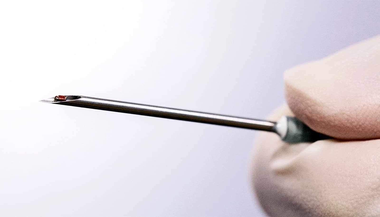
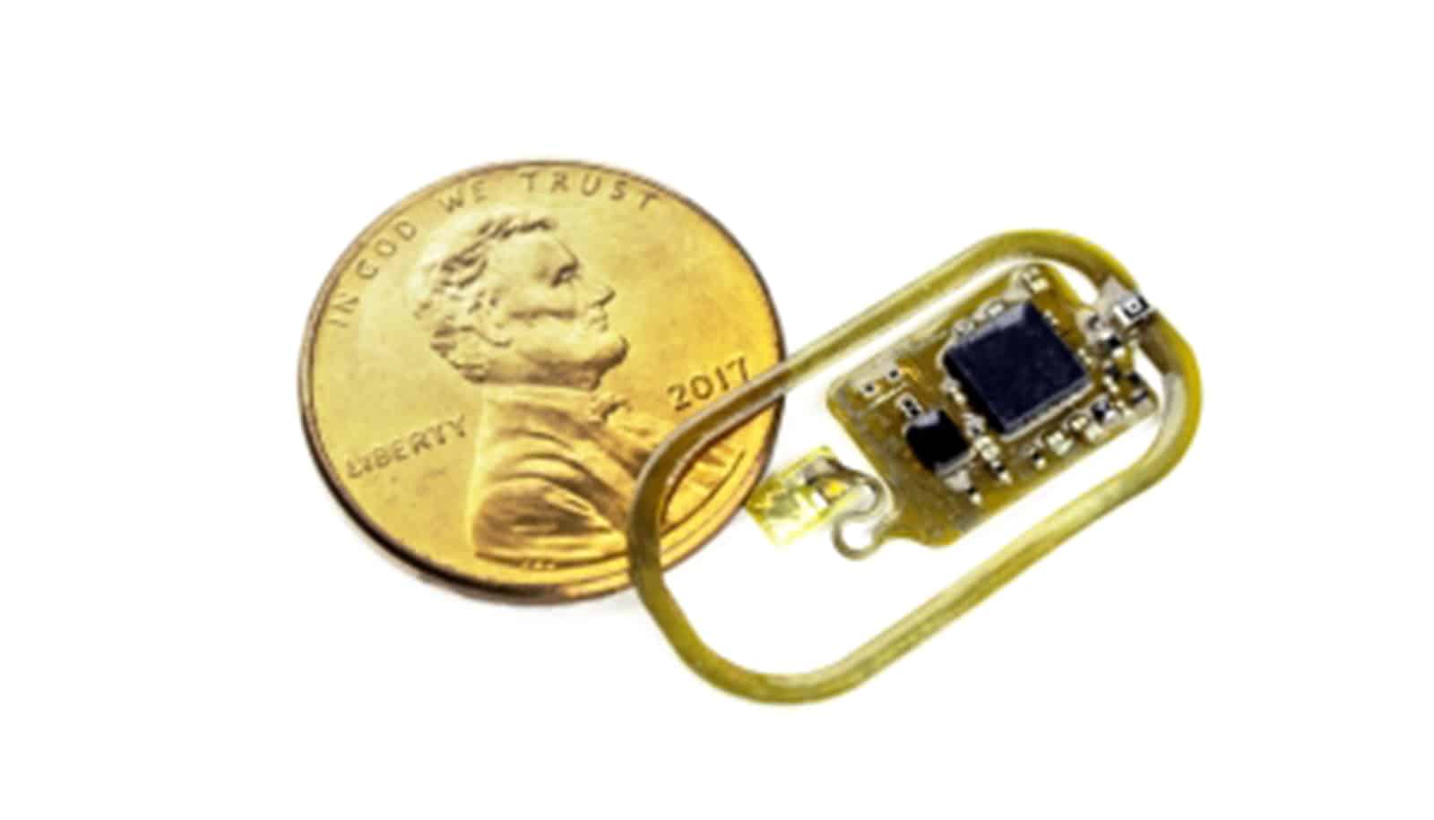
“The device’s thin structure, roughly as thick as a sheet of paper, means it can conform to the curvature of the bone, forming a tight interface,” says Alex Burton, a doctoral student in biomedical engineering and co-first author of the study. “They also do not need a battery. This is possible using a power casting and communication method called near-field communication, or NFC, which is also used in smartphones for contactless pay.”
The outer layers of bones shed and renew just like the outer layers of skin. So, if a traditional adhesive was used to attach something to the bone, it would fall off after just a few months. To address this challenge, coauthor John Szivek, a professor of orthopedic surgery and biomedical engineering and BIO5 Institute member, developed an adhesive that contains calcium particles with an atomic structure similar to bone cells, which is used as to secure osseosurface electronics to the bone.
“The bone basically thinks the device is part of it, and grows to the sensor itself,” Gutruf says. “This allows it to form a permanent bond to the bone and take measurements over long periods of time.”
For instance, a doctor could attach the device to a broken or fractured bone to monitor the healing process. This could be particularly helpful in patients with conditions such as osteoporosis, since they frequently suffer refractures. Knowing how quickly and how well the bone is healing could also inform clinical treatment decisions, such as when to remove temporary hardware like plates, rods, or screws.
Some patients are prescribed drugs designed to speed up bone healing or improve bone density, but these prescriptions can have side effects. Close bone monitoring would allow physicians to make more informed decisions about drug dosage levels.
Source: University of Arizona