Natural history museums have entered a new stage of scientific discovery and accessibility with the completion of a project to create 3D reconstructions of vertebrate specimens and make them freely available online.
Natural history museums got their start in the 16th century as cabinets of curiosity, in which a few wealthy individuals amassed rare and exotic specimens, which they kept mostly to themselves. Since then, museums have become a resource for the public, with exhibits that showcase biodiversity for anyone interested in learning about it.
However, most natural history collections remain behind closed doors, accessible only to scientists who must either travel to see them or ask that a small number of specimens be mailed on loan. The research team behind the project, open Vertebrate (oVert), wants to change that.

“If you require someone to get on a plane and travel to you to collaborate, that’s prohibitive in a lot of ways,” says David Blackburn, principal investigator of the oVert project and curator of herpetology at the Florida Museum of Natural History. “Now we have scientists, teachers, students, and artists around the world using these data remotely.”
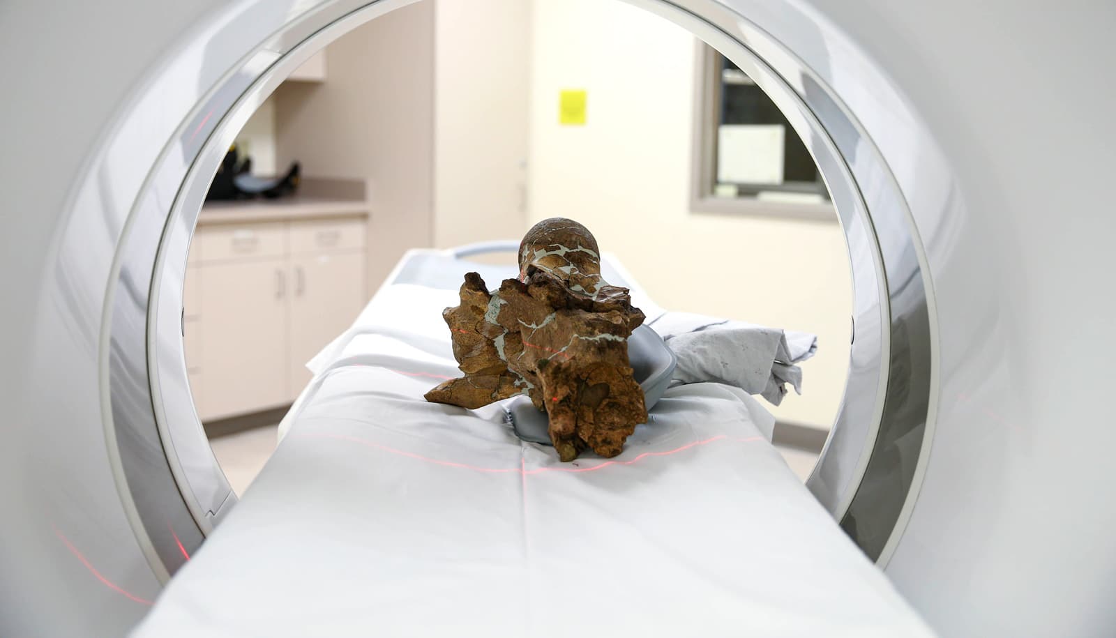
Between 2017 and 2023, oVert project members took CT scans of more than 13,000 specimens, with representative species across the vertebrate tree of life. This includes more than half the genera of all amphibians, reptiles, fish, and mammals.
More than 1,600 specimens from University of Michigan Museum of Zoology collections were scanned as part of the oVert project—mostly mammals, reptiles, amphibians, and fish, along with a few birds. The museum also scanned several hundred vertebrate specimens for partner institutions.

“The University of Michigan Museum of Zoology is the home to some of the largest collections of vertebrates in the world. We were able to utilize those collections and our own in-house CT scanner to produce 3D models of thousands of our voucher specimens, as well as material from collaborating collections,” says Cody Thompson, mammal collections manager and associate research scientist at the museum.
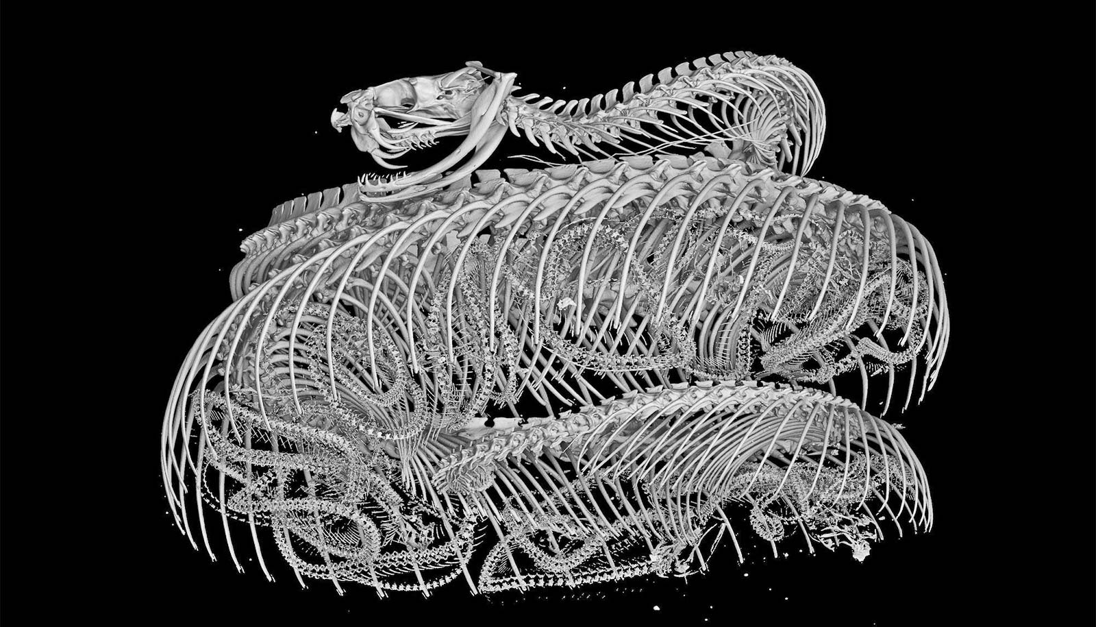
CT scanners use high-energy X-rays to peer past an organism’s exterior and view the dense bone structure beneath. Thus, skeletons make up the majority of oVert reconstructions. A small number of specimens were also stained with a temporary contrast-enhancing solution that allowed researchers to visualize soft tissues, such as skin, muscle, and other organs.
The models give an intimate look at internal portions of a vertebrate specimen that could previously only be observed through destructive dissection and tissue sampling.
“Museums are constantly engaged in a balancing act,” Blackburn says. “You want to protect specimens, but you also want to have people use them. oVert is a way of reducing the wear and tear on samples while also increasing access, and it’s the next logical step in the mission of museum collections.”
The project’s goal was to initially scan only specimens preserved in ethyl alcohol, which represent the bulk of fish, reptile, and amphibian collections.
Specimens that are too large for fluid preservation are also unable to fit into a CT scanner, but researchers were reluctant to leave these out. A partnering grant to the Idaho Museum of Natural History was used to create a digital model of a humpback whale.
The entire whale was too big to photograph with sufficient resolution, so researchers painstakingly took apart the skeleton, photographed each individual bone, then reassembled the physical and digital specimen.
Even moderately sized specimens at times required a little ingenuity, as was the case with a set of iconic tortoises at the California Academy of Sciences.
“They have the largest Galapagos tortoise collection in the world. These are not things you put in boxes and loan,” Blackburn says.
Using funds from another partnering grant, curatorial staff members had to come up with a way to photograph each tortoise in a 360-degree rotation. Photographing their undersides was problematic, as their curved shells made it impossible to keep them upright. After a few trial-and-error runs, they settled on placing the specimens on top of inflatable swimming tubes.
At the University of Michigan, several roadkill mammals, including a red fox and a porcupine, were among the animals scanned for the oVert project. Two recently deceased zoo animals—a polar bear and a tiger—were also scanned, with the help of the large-format CT scanner at Michigan State University’s veterinary college, Thompson says.
Scientists have already used data from the project to gain astonishing insights into the natural world.
A massive study of more than 500 oVert specimens revealed that frogs have lost and regained teeth more than 20 times throughout their evolutionary history. Another study concluded that Spinosaurus, a massive dinosaur that was larger than Tyrannosaurus rex and thought to be aquatic, would have actually been a poor swimmer.
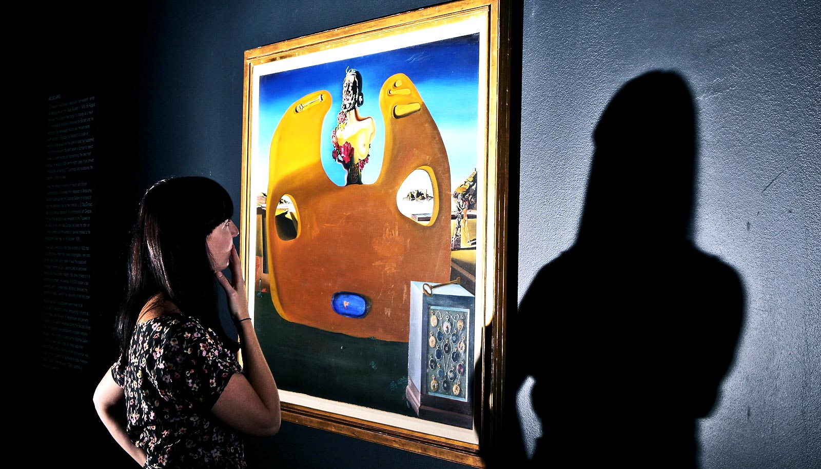
But the value of oVert extends beyond scientific inquiry. Artists have used the 3D models to create realistic animal replicas, photographs of oVert specimens have been displayed as museum exhibits, and specimens have been incorporated into virtual reality headsets that give users the chance to interact with and manipulate them.
oVert models have also been an invaluable resource for educators. From the outset of the project, Blackburn and his colleagues wanted to place a strong emphasis on K-12 outreach. They organized workshops where teachers could learn how to use the data in the classroom.
“It’s been a game-changer for my evolution unit,” says Jennifer Broo, a high school teacher in Cincinnati. “I teach juniors and seniors, and I absolutely love them, but they can be a tough audience. They know when things are fake, which makes them less engaged. Using the oVert models, you can teach concepts at an appropriate level while also maintaining the authenticity of the science. My class has gotten so much better because I have had the opportunities to work with and expose my students to real data.”
There’s virtually no end to the number of things oVert could be used for. The biggest challenge will be creating tools that are sophisticated enough to analyze the data. Never before have this many 3D natural history specimens been publicly available and instantly accessible, and it will take further developments in machine learning and supercomputing to use them to their full potential.
“One of the great things that oVert has done is rejuvenate research programs in anatomy and morphology,” says Thompson. “In a sort of serendipitous way, the COVID-19 pandemic provided the perfect backdrop for scientists to dig into the oVert datasets and push those sciences and related disciplines back to the forefront, which is great for natural history museums.”
Check out the library of 3D scans.
A paper detailing the project appears in the journal BioScience. The project is led by the Florida Museum of Natural History. Additional contributors to the project include the University of Michigan, the University of Florida, the University of Washington, and other institutions.
The National Science Foundation funded the project.
Source: Jerald Pinson for University of Michigan