A new simulation shows how blood flows through the heart.
This view inside a beating heart is one not seen—or even seeable—until now, according to Tayfun Tezduyar.
Tezduyar, professor of mechanical engineering at Rice University’s George R. Brown School of Engineering, says such visualizations can help clinicians understand the mechanisms that push blood through the body and fix things when they go awry.
Tezduyar is an expert in modeling interactions between fluids and structures whose work encompasses biomechanics, including the circulatory system, and large-scale systems like parachutes, wind turbines, and ground vehicles.
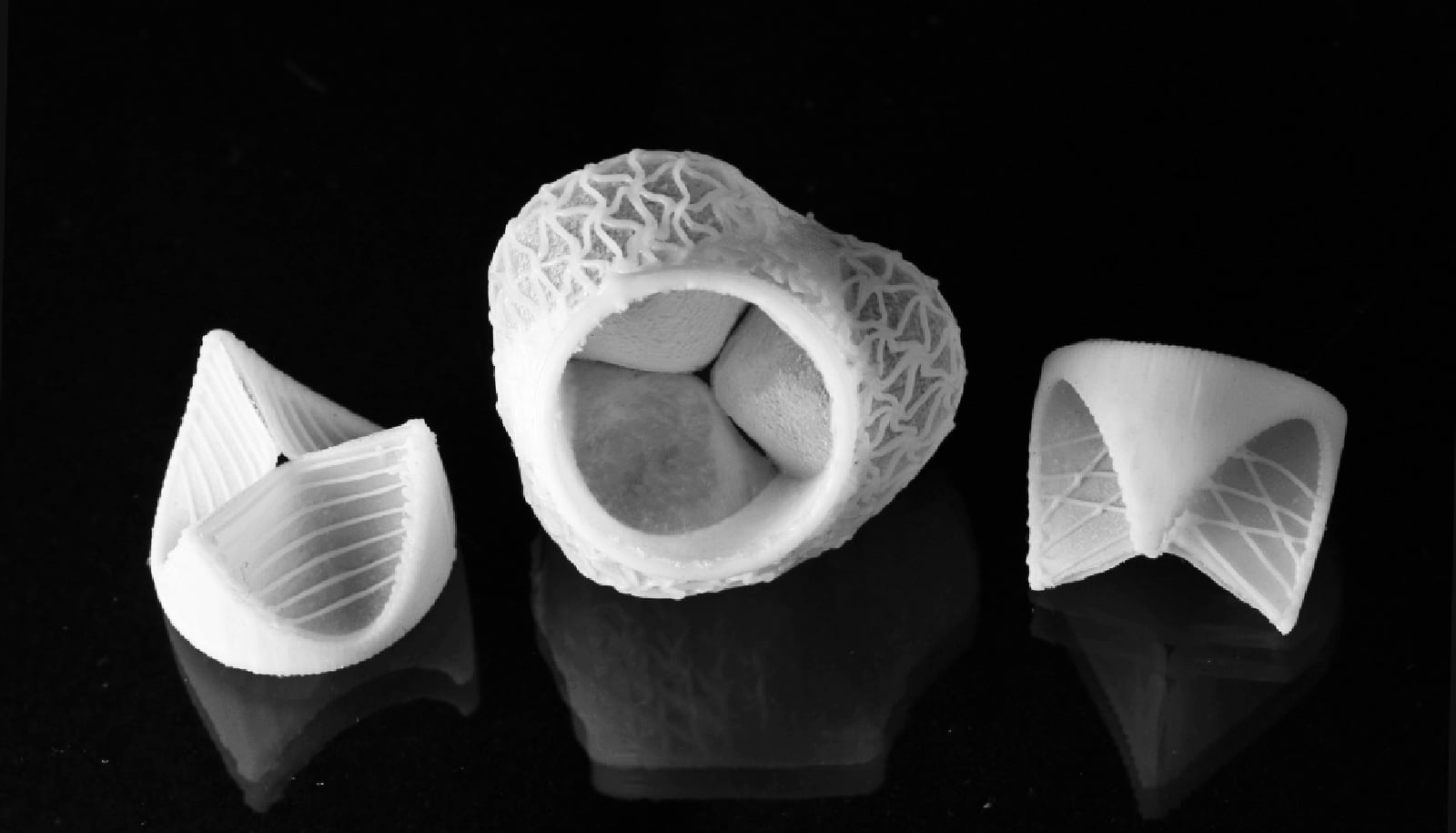
In this research, Tezduyar and his colleagues at Waseda University in Japan tackled what he says is one of the toughest problems in biomechanics: a way to accurately characterize the flow of blood around and through the heart’s valves, taking into account the flow details near the surfaces of the valve’s three leaflets along the way.
The animation shows two views of the ventricle-valve-aorta, top and side, and reveals in great detail how the ventricle fills with blood, how quickly the three-leaf valve opens to relieve the pressure, and the turbulent flow of blood as it pushes through.
“This is a computational technology that no one else in the world currently has with this level of accuracy near the leaflet surfaces,” Tezduyar says. “We’ve become aware recently that people at the Texas Medical Center are asking for CFD (computational fluid dynamics) simulations of the cardiovascular system. I think perhaps they meant what happens just in the aorta, and that would have been a lot easier. But we went a little beyond.”
Tezduyar says the simulation incorporates the valve mechanism and fluid flow through the aorta and its three branch arteries.
“That’s a very difficult computation,” he says. “When someone needs to make decisions based on a simulation, the main issue is how accurate and reliable it is. The opening and closing of the valves make it extremely complex, and generally the more complex a simulation becomes, the less reliable it could be.
“What we’ve done is keep both the complexity and accuracy,” he says. “It was painstaking work. Modeling three-leaflet valves that touch each other, fully close off blood flow and then open again, and doing that with accurate representation of the near-leaflet flow patterns, is a new and challenging class of problems in fluid mechanics.”
He says the model is detailed enough to capture the spiraling motion of blood in the aorta.
“We need to represent the flow patterns on these walls correctly to evaluate shear stress on the arterial walls, which is connected to the risk of aneurisms,” says Tezduyar, who co-leads the Team for Advanced Flow Simulation and Modeling with Kenji Takizawa, a professor in the School of Creative Science and Engineering at Waseda University.
“This is valuable information for doctors looking for ways to cure heart disease,” he says. “For instance, the model would help them evaluate replacement valves, including the geometry, thickness, and material.”
The simulation of a working ventricle-valve-aorta represents calculations first presented by Tezduyar and his Waseda colleagues in 2020.
Takizawa was a research associate at Rice beginning in 2007. His research associate, Takuya Terahara, who also spent time at Rice as a visiting PhD student in 2018, produced the animation. Tezduyar says he and Takizawa are amenable to discussing with doctors long-term collaborative work on patient-specific simulations.
Source: Rice University