Biologists have grown human retina tissue from scratch to learn how the cells that let us see in color form.
The work may lay the groundwork for therapies for eye diseases such as color blindness and macular degeneration. It also establishes lab-created “organoids”—artificially grown organ tissue—as a model to study human development on a cellular level.
“Everything we examine [in a retina organoid] looks like a normal developing eye, just growing in a dish,” says Robert Johnston, a developmental biologist at Johns Hopkins University. “You have a model system that you can manipulate without studying humans directly.”
The fate of stem cells
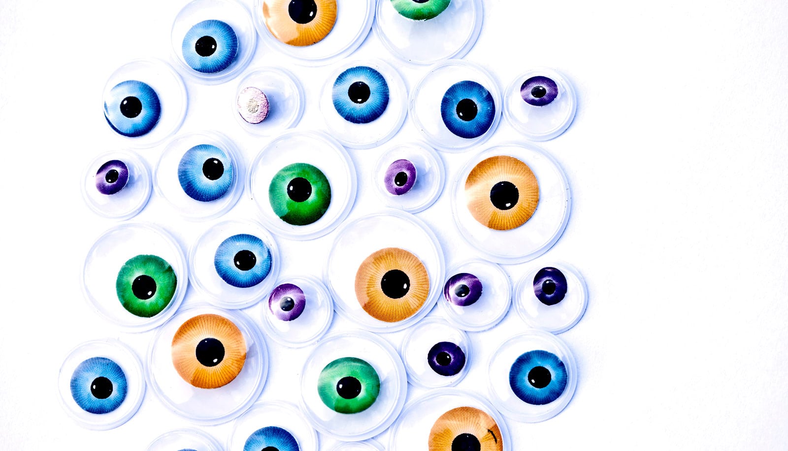
Johnston’s lab explores how a cell’s fate is determined—what happens in the womb to turn a developing stem cell into a cell with a specific function. In the retina research, he and his team focused on the development of cells that allow people to see blue, red, and green—the three cone photoreceptors in the human eye.
While most vision research is done on mice and fish, neither of those species has the dynamic daytime and color vision of humans. So Johnston’s team created the human eye tissue they needed from stem cells.
“Trichromatic color vision differentiates us from most other mammals,” says lead author Kiara Eldred, a graduate student. “Our research is really trying to figure out what pathways these cells take to give us that special color vision.”
Over months, as the cells grew in the lab and became full-blown retina tissue, the team found the blue-detecting cells materialized first, followed by the red- and green-detecting ones. In both cases, they found, the key to the molecular switch was the ebb and flow of thyroid hormone. Importantly, the thyroid gland, which of course wasn’t in the lab dish, didn’t control the level of this hormone, but the eye tissue itself did.
Once the researchers understood how the amount of thyroid hormone dictated whether the cells became blue or red and green receptors, they could manipulate the outcome, creating retinas that—if they had been part of a complete human eye—would have seen only blue, and others that would have detected green and red.
Insight into vision
The finding that thyroid hormone is essential for creating red-green cones provides insight into why pre-term babies, who have lowered thyroid hormone levels as they are lacking the maternal supply, have a higher incidence of vision disorders.
“If we can answer what leads a cell to its terminal fate, we are closer to being able to restore color vision for people who have damaged photoreceptors,” Eldred says. “This is a really beautiful question, both visually and intellectually—what is it that allows us to see color?”
These findings are a first step for the lab. In the future, the researchers would like to use organoids to learn even more about color vision and the mechanisms involved in the creation of other regions of the retina, such as the macula. Since macular degeneration is one of the leading causes of blindness in people, understanding how to grow a new macula could lead to clinical treatments.
“What’s exciting about this is our work establishes human organoids as a model system to study mechanisms of human development,” Johnston says. “What’s really pushing the limit here is that these organoids take nine months to develop just like a human baby. So what we’re really studying is fetal development.”
The research appears in the journal Science.
Additional researchers who contributed to the work are from Johns Hopkins; the Shiley Eye Institute of the University of California, San Diego; and the National Institutes of Health.
The Pew Charitable Trusts, the Howard Hughes Medical Institute, the National Science Foundation, and the National Institutes of Health funded the study.
Source: Johns Hopkins University