Researchers have cracked the longstanding mystery of how groups of cells manage to build human tissues and organs during the embryo stage.
Not only does it bring a century-old hypothesis into the modern age, the study and its techniques provide the researchers a foundation to study other questions key to human health, such as how cancers form and spread and how to engineer organs.
“In a nutshell, we discovered a fundamental physical mechanism that cells use to mold embryonic tissues into their functional 3D shapes,” says Otger Campàs, a professor of mechanical engineering in the College of Engineering at the University of California, Santa Barbara, and chair in systems biology. His group investigates how living systems organize themselves to build the remarkable structures and shapes found in nature.
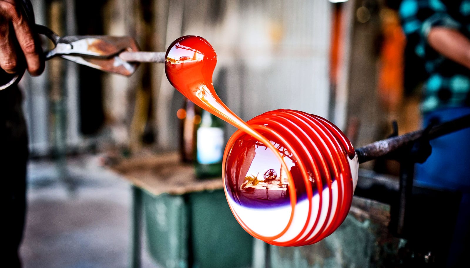
Cells coordinate by exchanging biochemical signals, but they also hold to and push on each other to build the body structures we need to live, such as the eyes, lungs, and heart. And, as it turns out, sculpting the embryo is not far from glass molding or 3D printing.
In their new work, which appears in the journal Nature, Campàs and colleagues show how cell collectives switch from fluid to solid states in a controlled manner to build the vertebrate embryo, in a way similar to how glass is molded into vases or how items are 3D printed. Or, if you like, we 3D print ourselves, from the inside.
From fluid to solid
Most objects begin as fluids. From metallic structures to gelatin desserts, their shape is made by pouring the molten original materials into molds, then cooling them to get the solid objects we use.
As in a Chihuly glass sculpture, made by carefully melting portions of glass to slowly reshape it into life, cells in certain regions of the embryo are more active and “melt” the tissue into a fluid state that can be restructured. Once done, cells “cool down” to settle the tissue shape, Campàs explains.
“The transition from fluid to solid tissue states that we observed is known in physics as ‘jamming’,” Campàs says. “Jamming transitions are a very general phenomena that happens when particles in disordered systems, such as foams, emulsions, or glasses, are forced together or cooled down.”
Techniques Campàs and his group previously developed to measure the forces between cells inside embryos, and also to exert miniscule forces on the cells as they build tissues and organs, enabled this discovery.
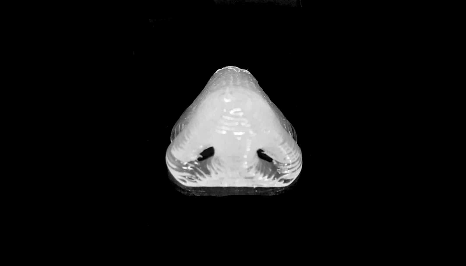
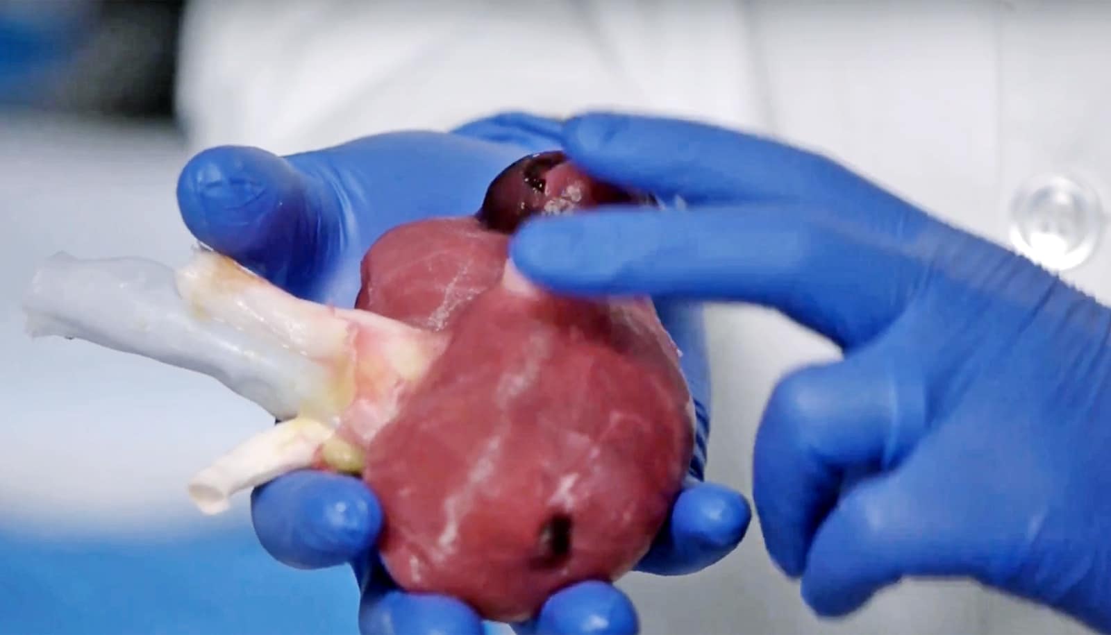
Using zebrafish embryos, favored for their optical transparency but developing much like their human counterparts, the researchers placed tiny droplets of a specially engineered ferromagnetic fluid between the cells of the growing tissue. The spherical droplets deform as the cells around them push and pull, allowing researchers to see the forces that cells apply on each other. And, by making these droplets magnetic, they also could exert tiny stresses on surrounding cells to see how the tissue would respond.
“We were able to measure physical quantities that couldn’t be measured before, due to the challenge of inserting miniaturized probes in tiny developing embryos,” says lead author Alessandro Mongera, a postdoctoral fellow.
“Zebrafish, like other vertebrates, start off from a largely shapeless bunch of cells and need to transform the body into an elongated shape, with the head at one end and tail at the other,” Campàs says.
The physical reorganization of the cells behind this process had always been something of a mystery. Surprisingly, researchers found that the cell collectives making the tissue were physically like a foam (as in beer froth) that jammed during development to ‘freeze’ the tissue architecture and set its shape.
100-year-old idea
These observations confirm a remarkable intuition Victorian-era Scottish mathematician D’Arcy Thompson made 100 years ago in his “On Growth and Form.”
“He was convinced that some of the physical mechanisms that give shapes to inert materials were also at play to shape living organisms. Remarkably, he compared groups of cells to foams and even the shaping of cells and tissues to glassblowing,” Campàs says.
A century ago, there were no instruments that could directly test the ideas Thompson proposed, Campàs adds, though Thompson’s work continues to be cited to this day.
The new paper also provides a jumping-off point from which Campàs and colleagues can begin to address other processes of embryonic development and related fields, such as how tumors physically invade surrounding tissues and how to engineer organs with specific 3D shapes.
“One of the hallmarks of cancer is the transition between two different tissue architectures. This transition can in principle be explained as an anomalous switch from a solid-like to a fluid-like tissue state,” Mongera explains. “The present study can help elucidate the mechanisms underlying this switch and highlight some of the potential druggable targets to hinder it.”