Artificial intelligence can improve the quality of optoacoustic imaging, researchers report.
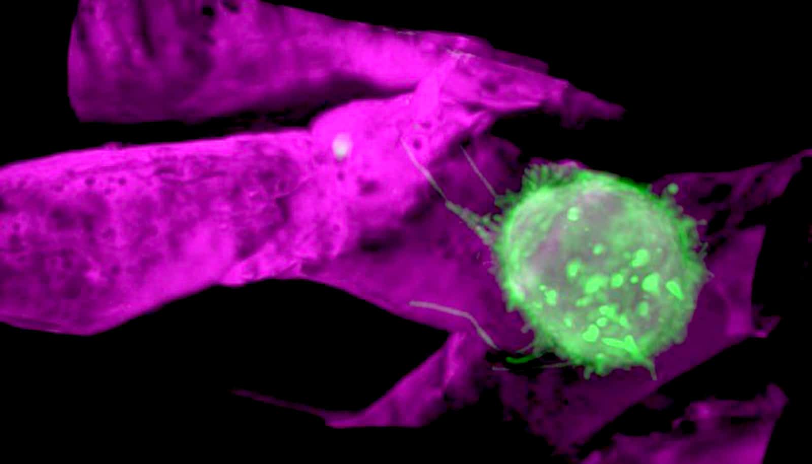
This relatively young medical imaging technique can be used for applications such as visualizing blood vessels, studying brain activity, characterizing skin lesions, and diagnosing breast cancer. However, quality of the rendered images is very dependent on the number and distribution of sensors used by the device: the more of them, the better the image quality.
The new approach allows for substantial reduction of the number of sensors without giving up on the resulting image quality. This makes it possible to reduce the device cost, increase imaging speed, or improve diagnosis.
What is optoacoustic imaging?
Optoacoustics is similar in some respects to ultrasound imaging. In the latter, a probe sends ultrasonic waves into the body, which the tissue reflects. Sensors in the probe detect the returning sound waves and generate a picture of the inside of the body. Optoacoustic imaging instead sends very short laser pulses into the tissue, where they’re absorbed and converted into ultrasonic waves. Similarly to ultrasound imaging, researchers can then detect the waves convert them into images.
The team searched for a way to enhance image quality of low-cost optoacoustic devices that possess only a small number of ultrasonic sensors.
To do this, they started off by using a self-developed high-end optoacoustic scanner with 512 sensors, which delivered superior-quality images. They had an artificial neural network analyze the pictures, and it learned the features of the high-quality images.
Next, the researchers discarded the majority of the sensors, so that only 128 or 32 sensors remained, with a detrimental effect on the image quality. Due to the lack of data, distortions known as streak type artifacts appeared in the images. It turned out, however, that the previously trained neural network could largely correct for these distortions, thus bringing the image quality closer to the measurements obtained with all the 512 sensors.
In optoacoustics, the image quality increases not only with the number of sensors researchers use, but also when they capture information from as many directions as possible: the larger the sector in which researchers arrange the sensors around the object, the better the quality. The machine learning algorithm was also successful in improving quality of images that researchers recorded from just a narrowly circumscribed sector.
“This is particularly important for clinical applications, as the laser pulses cannot penetrate the entire human body, hence the imaged region is normally only accessible from one direction,” according to Daniel Razansky, professor of biomedical imaging at the University of Zurich and ETH Zurich.
AI’s helping hand
The scientists stress that their approach is not limited to optoacoustic imaging. Because the method operates on the reconstructed images, not the raw recorded data, it is also applicable to other imaging techniques.
“You can basically use the same methodology to produce high-quality images from any sort of sparse data,” Razansky says. He explains that physicians often face the challenge of interpreting poor quality images from patients. “We show that such images can be improved with AI methods, making it easier to attain more accurate diagnosis.”
For Razansky, this research work is a good example of how scientists can use existing methods of artificial intelligence.
“Many people think that AI could replace human intelligence. This is probably exaggerated, at least for the currently available AI technology,” he says. “It can’t replace human creativity, yet may release us from some laborious, repetitive tasks.”
In their current research, the scientists used an optoacoustic tomography device customized for small animals, and trained the machine learning algorithms with images from mice. The next step will be to apply the method to optoacoustic images from human patients, Razansky says.
No contrast agents necessary
Unlike optoacoustics (also known as photoacoustics), many imaging techniques, such as ultrasound, X-ray, or MRI, are mainly suitable for visualizing anatomical alterations in the body. To receive additional functional information, for instance concerning blood flow or metabolic changes, doctors have to administer contrast agents or radioactive tracers to the patient before the imaging.
In contrast, the optoacoustic method can visualize functional and molecular information without introducing contrast agents. One example is local changes in tissue oxygenation—an important landmark of cancer that can be useful for early diagnosis. Lipid content in blood vessels is yet another potential disease marker, which can aid an early detection of cardiovascular diseases.
It should be noted, however, that because the light waves used in optoacoustic imaging, unlike other waves, do not fully penetrate the human body, the method is only suitable for investigating tissues to a depth of a few centimeters (a little more than an inch) beneath the skin.
The research appears in Nature Machine Intelligence.
Source: ETH Zurich