Scientists have developed a new cell imaging technology that creates high-resolution “movies” of cells in their 3D environment and captures subcellular processes.
Published in Science, the research reveals a technology that shows the phenotypic diversity within cells across different organisms and developmental stages and in conditions such as mitosis, immune processes, and in metastases.
The technique, which combines lattice light sheet microscopy (LLSM) and adaptive optics (AO), offers scientists investigating cancer and other diseases new insights into how cells operate and adapt to different physiological environments.
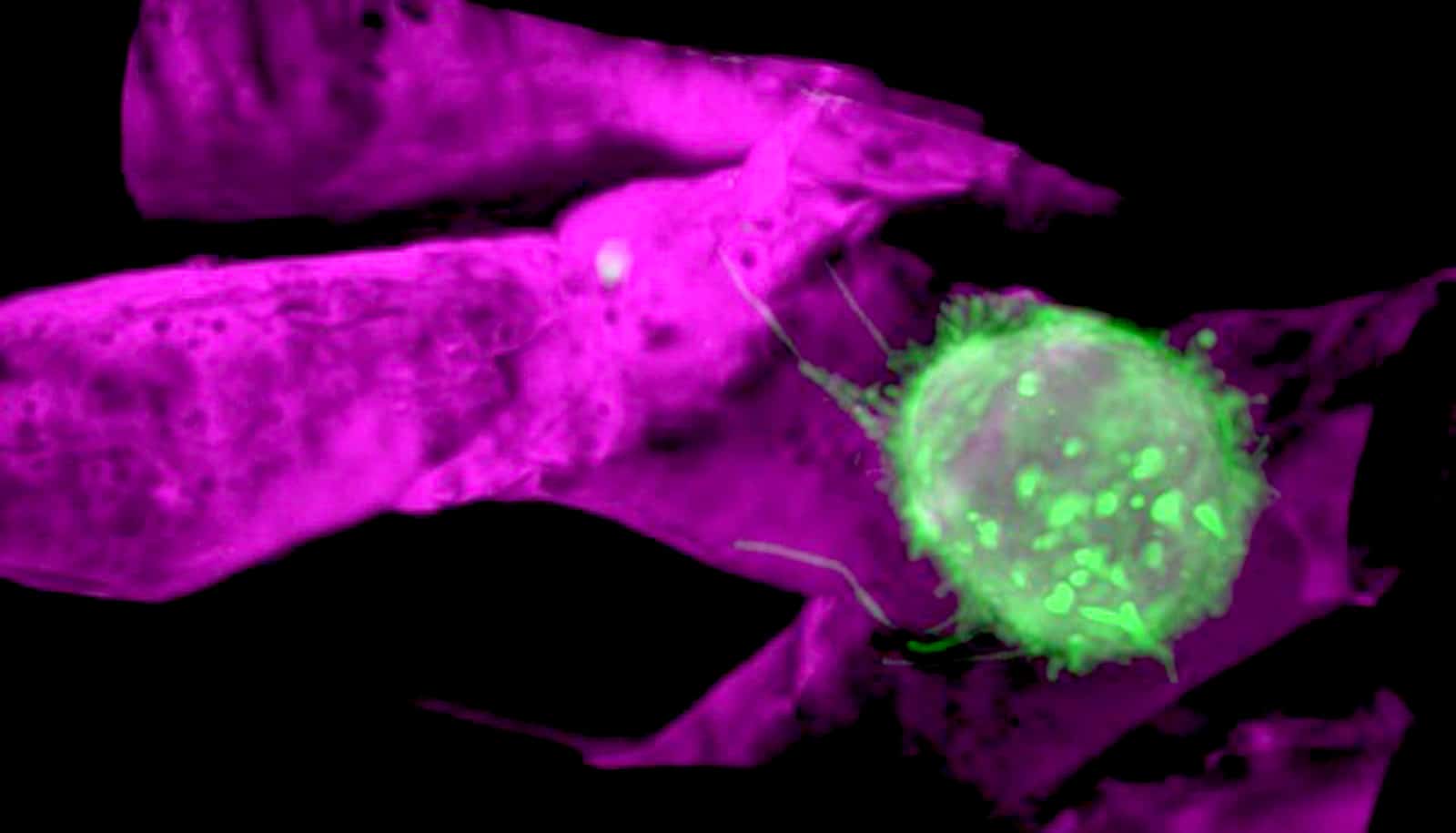
In the paper, professors David Q. Matus and Benjamin L. Martin, both of the Stony Brook University Cancer Center and department of biochemistry and cell biology, used AO-LLSM to capture and visualize the behavior of human breast cancer cells injected into zebrafish vasculature.
Scientists map path into cell’s nucleus
They successfully captured time-lapse movies at high resolution of breast cancer cells mimicking the cell behaviors characteristic of immune cells (leukocytes). These behaviors include rolling, crawling, and invading out of the vasculature. See the cells moving in the video above.
“By observing and characterizing these behaviors, such as cancer cells adopting leukocyte-like behaviors, we may be able to discover new avenues to target the spread or dissemination of metastatic cancer cells,” says Matus.
The National Science Foundation, the National Institutes of Health, the Damon Runyon Cancer Research Foundation, and the Carol M. Baldwin Breast Cancer Research Fund supported the work.
Source: Stony Brook University