Through long shifts at the helm of a highly sophisticated microscope, researchers have recorded reactions at near-atomic-scale resolution. The work could someday help our phone batteries last longer and our electric vehicles go farther on a single charge.
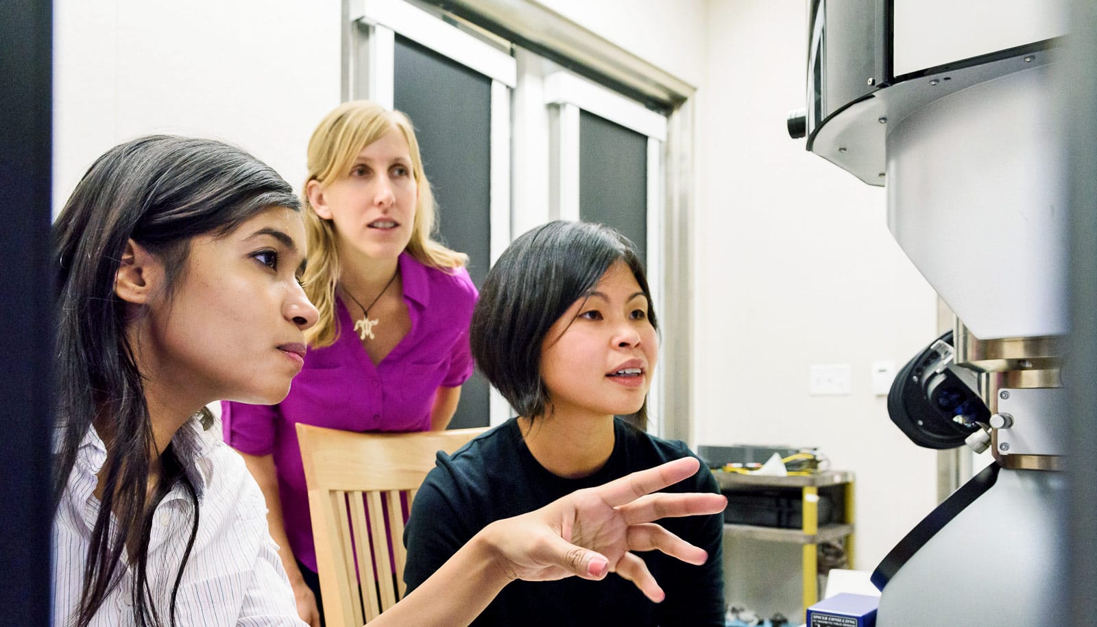
In a lab 18 feet underground, researchers in the Dionne lab at Stanford University conducted the arduous experiments—sometimes requiring 30 continuous hours of work—to capture real-time, dynamic visualizations of atoms moving in and out of nanoparticles less than 100 nanometers in size, with a resolution approaching 1 nanometer.
“The ability to directly visualize reactions in real time with such high resolution will allow us to explore many unanswered questions in the chemical and physical sciences,” says Jen Dionne, associate professor of materials science and engineering and senior author of the paper about the work in Nature Communications. “While the experiments are not easy, they would not be possible without the remarkable advances in electron microscopy from the past decade.”
Watch nanotubes wriggle to form a bridge
Their experiments focused on hydrogen moving into palladium, a class of reactions known as an intercalation-driven phase transition. This reaction is physically analogous to how ions flow through a battery or fuel cell during charging and discharging. Observing this process in real time provides insight into why nanoparticles make better electrodes than bulk materials and fits into Dionne’s larger interest in energy storage devices that can charge faster, hold more energy and stave off permanent failure.
A ‘ghost’ in the joystick
For these experiments, the Dionne lab created palladium nanocubes, a form of nanoparticle that ranged in size from about 15 to 80 nanometers, and then placed them in a hydrogen gas environment within an electron microscope. The researchers knew that hydrogen would change both the dimensions of the lattice and the electronic properties of the nanoparticle. They thought that, with the appropriate microscope lens and aperture configuration, techniques called scanning transmission electron microscopy and electron energy loss spectroscopy might show hydrogen uptake in real time.
After months of trial and error, the results were extremely detailed, real-time videos of the changes in the particle as hydrogen was introduced. The entire process was so complicated and novel that the first time it worked, the lab didn’t even have the video software running, leading them to capture their first movie success on a smartphone.
Following these videos, they examined the nanocubes during intermediate stages of hydrogenation using a second technique in the microscope, called dark-field imaging, which relies on scattered electrons. In order to pause the hydrogenation process, the researchers plunged the nanocubes into an ice bath of liquid nitrogen mid-reaction, dropping their temperature to 100 degrees Kelvin (-280 F). These dark-field images served as a way to check that the application of the electron beam hadn’t influenced the previous observations and allowed the researchers to see detailed structural changes during the reaction.
Team measures nanoparticles with microscope’s tiny tip
“With the average experiment spanning about 24 hours at this low temperature, we faced many instrument problems and called Ai Leen Koh [coauthor and research scientist at Stanford’s Nano Shared Facilities] at the weirdest hours of the night,” recalls Fariah Hayee, co-lead author of the study and graduate student in the Dionne lab. “We even encountered a ‘ghost-of-the-joystick problem,’ where the joystick seemed to move the sample uncontrollably for some time.”
While most electron microscopes operate with the specimen held in a vacuum, the microscope used for this research has the advanced ability to allow the researchers to introduce liquids or gases to their specimen.
“Without these specific tools, we wouldn’t be able to introduce hydrogen gas or cool down our samples enough to see these processes take place,” says Tarun Narayan, co-lead author of the study and recent doctoral graduate from the Dionne lab.
Self-healing
Aside from being a widely applicable proof of concept for this suite of visualization techniques, watching the atoms move provides greater validation for the high hopes many scientists have for nanoparticle energy storage technologies.
This crystal heals itself after cracking in two
The researchers saw the atoms move in through the corners of the nanocube and observed the formation of various imperfections within the particle as hydrogen moved within it. This sounds like an argument against the promise of nanoparticles but that’s because it’s not the whole story.
“The nanoparticle has the ability to self-heal,” says Dionne. “When you first introduce hydrogen, the particle deforms and loses its perfect crystallinity. But once the particle has absorbed as much hydrogen as it can, it transforms itself back to a perfect crystal again.”
The researchers describe this as imperfections being “pushed out” of the nanoparticle. This ability of the nanocube to self-heal makes it more durable, a key property needed for energy storage materials that can sustain many charge and discharge cycles.
Next steps
As the efficiency of renewable energy generation increases, the need for higher quality energy storage is more pressing than ever. It’s likely that the future of storage will rely on new chemistries and the findings of this research, including the microscopy techniques the researchers refined along the way, will apply to nearly any solution in those categories.
The team could next look at a variety of material compositions, or compare how the sizes and shapes of nanoparticles affect the way they work, and, soon, take advantage of new upgrades to their microscope to study light-driven reactions. At present, Hayee has moved on to experimenting with nanorods, which have more surface area for the ions to move through, promising potentially even faster kinetics.
Funding came from the Air Force Office of Scientific Research, the National Science Foundation, the SLAC National Accelerator Laboratory in concert with the Department of Energy, the Foundation for Fundamental Research on Matter, and the Department of Energy Office of Science Graduate Fellowship Program.
Source: Stanford University