A new method can examine protein assembly in real time, in living cells, to find problems in the process and diagnose the resulting diseases, according to new research.
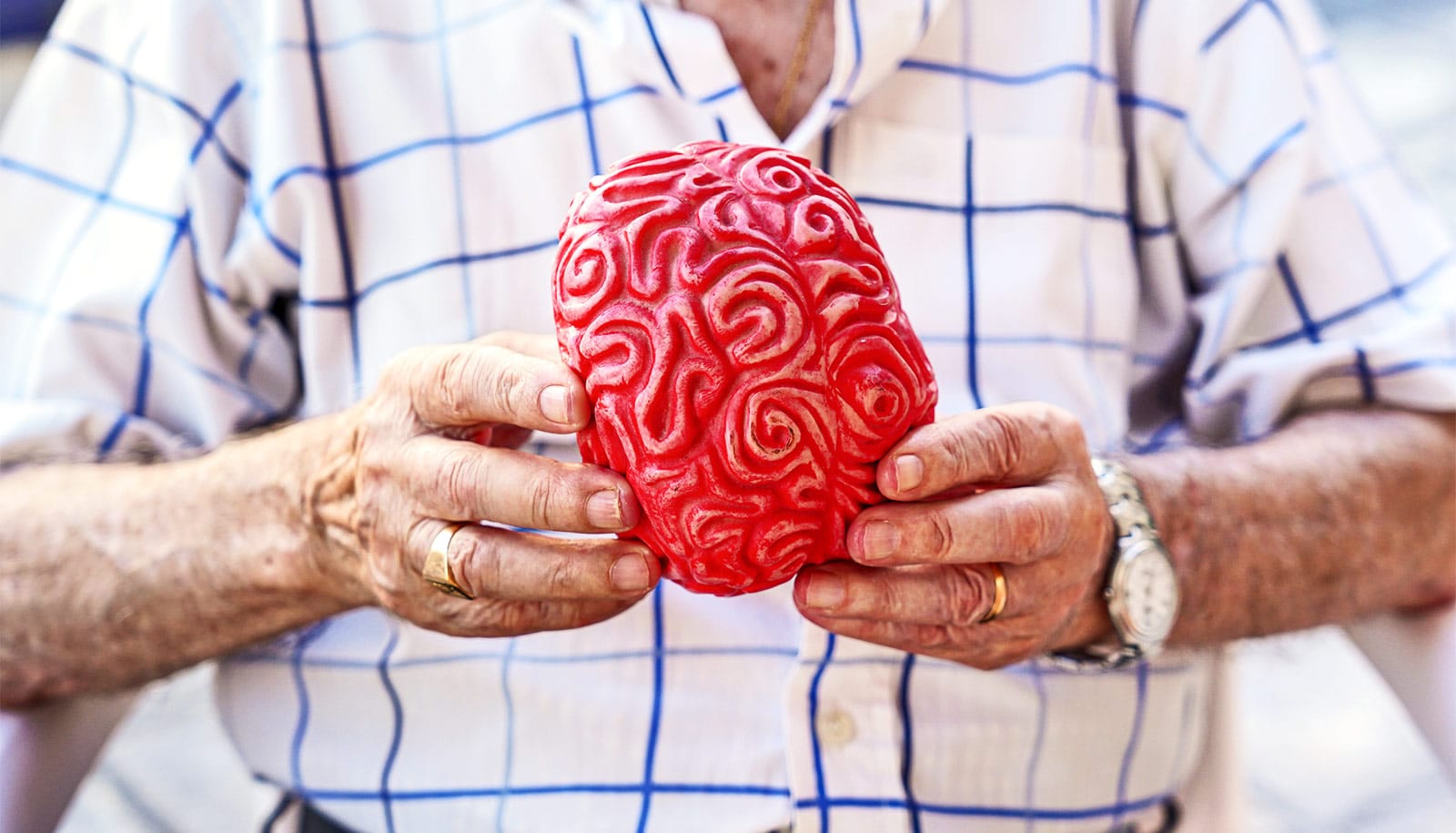
Proteins in the body need to be perfectly arranged, or folded, to do their jobs. When there is a misfolding, a number of problems and diseases—including diseases of aging and different cancers—can result.
“Normally this is done in test tubes…”
Proteins are organic molecules made up of different combinations of 20 amino acids, the building blocks of life. For any given protein, the amino acids must be joined together by chemical bonds and then folded properly, in single copy—called a monomer—or multiple copies, called an oligomer, to execute the protein’s function within cells.
Nearly a third of the proteins in the human body are assembled as oligomers to perform their biological tasks. Haemoglobin, for example, must be assembled as a tetramer (a specific type of oligomer with four units) to most efficiently transport oxygen through the blood stream.
A disruption of hemoglobin’s assembly would compromise the uptake of oxygen into cells, potentially causing damage to cells throughout the body due to lack of oxygen. That’s where the new study comes in.
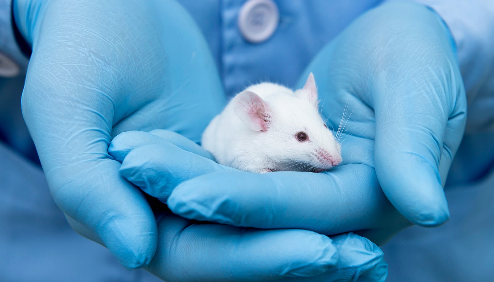
“We developed a novel method to examine protein assembly in real time and to dissect the structure-function relations in living cells. Normally this is done in test tubes,” says Yubin Zhou, associate professor at the Institute of Biosciences and Technology at Texas A&M University. “Our study meets the urgent and critical need for quantitative assessment of protein structure and monitoring of protein actions under native conditions.”
Zhou calls the engineered, genetically encoded mini-tags MoTags, which is short for monomer/oligomer detection tag. They are able to tell how many units are in the protein, which the researchers can then compare to how many there are supposed to be in that particular protein—and therefore determine if there is any misfolding.
It is thought that misfolding of proteins, which naturally happens during the aging process as proteins are damaged, can cause some of the major diseases associated with aging, from cancer to Alzheimer’s and Parkinson’s. Therefore, a method that can quickly tell if a protein is inappropriately assembled in real time, in living cells, will not only help scientists to better understand the pathogenic mechanisms, but also aid the diagnosis of disease and possible treatments.
This method can be repurposed to screen for drugs that can correct the misfolding or mis-assembly of disease-causing or disease-associated key proteins.
“In addition to protein structure-function relationship studies, our approach can detect protein to protein, protein to DNA or protein ligand to drug interactions,” says Guolin Ma, a postdoctoral fellow in Zhou’s lab who spearheaded the work. “Therefore, the live-cell screening assays against these interactions can be set up based on our method.”
How ‘machine’ untangles messed up proteins to save cells
“Our method can be widely adopted by any laboratories equipped with a standard fluorescence microscope to study protein chemistry, and thus is suitable for a broad application in the fields of protein chemistry, chemical biology, biochemistry research, as well as the development of assays to aid drug screening and therapeutic development in the near future,” says Yun Nancy Huang, also an assistant professor at the Texas A&M’s Institute of Biosciences and Technology.
“Our simple methods have straightforward readouts and circumvent laborious protein expression and purification procedures. We are thrilled the broad adaptability and wide applications of MoTags in biomedical research, as well as the potential for transforming the way biochemists to study their pet proteins.”
The research appears in Chemical Science.
Source: Texas A&M University