While a brain injury can be difficult to locate, a single region of the brain may show the impact of a concussion or repeated hits to the head, according to new research.
The finding, which appears in Science Advances, also supports the emerging idea that traumatic brain injury is not limited to people who sustain a concussion; it can result from repetitive head hits that are clinically silent—those that do not produce the visible signs or symptoms of a concussion. These subconcussive hits have been increasingly recognized as a potential threat to long-term brain health and as a possible cause of chronic traumatic encephalopathy (CTE).
The location of a brain injury varies widely from person to person, says coauthor Jeffrey Bazarian, professor of emergency medicine, neurology, neurosurgery, and public health sciences at the University of Rochester Medical Center. This is a major obstacle for physicians trying to diagnose brain injury using imaging techniques.
“This study is important because we found that no matter where the head gets hit, the force is translated into a single region of the brain known as the midbrain,” notes Bazarian, who treats concussion patients and conducts research related to traumatic brain injury. “Midbrain imaging might be a way in the future to diagnose injury from a single concussive head hit, as well as from repetitive sub-concussive head hits.”
‘Silent brain injuries’
“Our findings do not dispute the fact that head-injury effects are distributed throughout the brain, but the midbrain may serve as a ‘canary in a coal mine’ in terms of identifying damage,” says first author Adnan Hirad, a fourth-year medical student.
“From this study we know that the midbrain region, which is linked to brain functions often affected by a concussion, is the place to look to identify the impact of clinically defined concussions with visible symptoms and silent brain injuries that can’t be observed simply by looking at or behaviorally testing a player, on or off the field,” Hirad says.
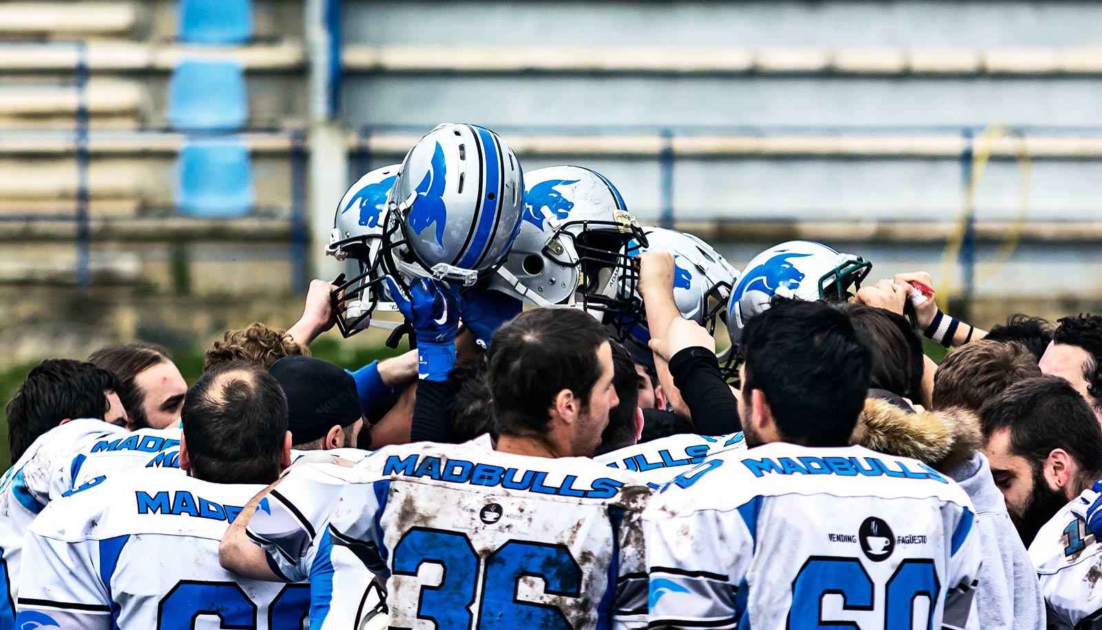
Researchers collected and analyzed data from 38 University of Rochester football players before and after three consecutive football seasons for the study. The researchers scanned the players’ brains in an MRI machine before and after a season of play, and equipped the football helmets they wore throughout the season with impact sensors that captured all hits above 10g force sustained during practices and games. Race car drivers feel the effects of 6 gs, and car crashes can produce brief forces of more than 100 gs.
Pinpointing the midbrain
The analysis showed a significant decrease in the integrity of the midbrain white matter following just one season of football as compared to the preseason. While only two players had clinically diagnosed concussions during the time they were followed in the study, the comparison of the post- and pre-season MRIs showed more than two-thirds of the players experienced a decrease in the structural integrity of their brain.
The research team also found that the amount of white matter damage correlated with the number of hits to the head players sustained. They also found that rotational acceleration (when the head twists from side to side or front to back) was linked more strongly and more consistently to changes in white matter integrity than linear acceleration (head-on impact).
“Public perception is that the big hits are the only ones that matter. It’s what people talk about and what we often see being replayed on TV,” says senior study author Brad Mahon, an associate professor of psychology at Carnegie Mellon University and scientific director of the Program for Translational Brain Mapping at the University of Rochester. “The big hits are definitely bad, but the public is likely missing what’s causing the long-term damage in players’ brains. It’s not just the concussions. It’s everyday hits, too. And the place to look for the effect of such hits, our study suggests, is the midbrain.”
The study’s authors looked for brain injury in the midbrain region because:
- It controls several brain functions that are frequently affected after concussion such as visual tracking, balance, auditory processing, and autonomic function.
- Prior research involving football players and boxers revealed that the highest strains following a concussive impact were concentrated in the midbrain.
- Published studies of concussive injuries in humans and animals also demonstrated that the midbrain is an important site of acute brain injury, as well as CTE.
This study is part of the newly launched Open Brain Project, a digital platform for exploration of the human brain and how it responds to and recovers from injury. Visitors to the site can explore models of the head hits sustained by football players in this study, and researchers can contribute their own datasets and post publications and commentary on a variety of topics covering brain tumors, epilepsy, stroke, and traumatic brain injury.
The University of Rochester Clinical and Translational Science Institute, the National Institutes of Health, and National Football League Charities funded the study.


