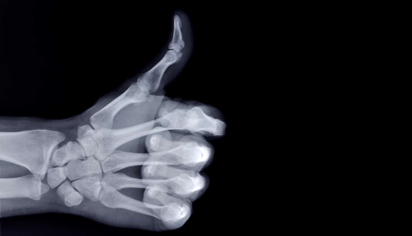
A new X-ray method has allowed researchers to identify zinc-sulfide nanoparticles in the timbers of the Mary Rose, the crown jewel of Henry VIII’s 16th century fleet, which are contributing to the ship’s decay.
Until now, this information was impossible to learn from the wood.
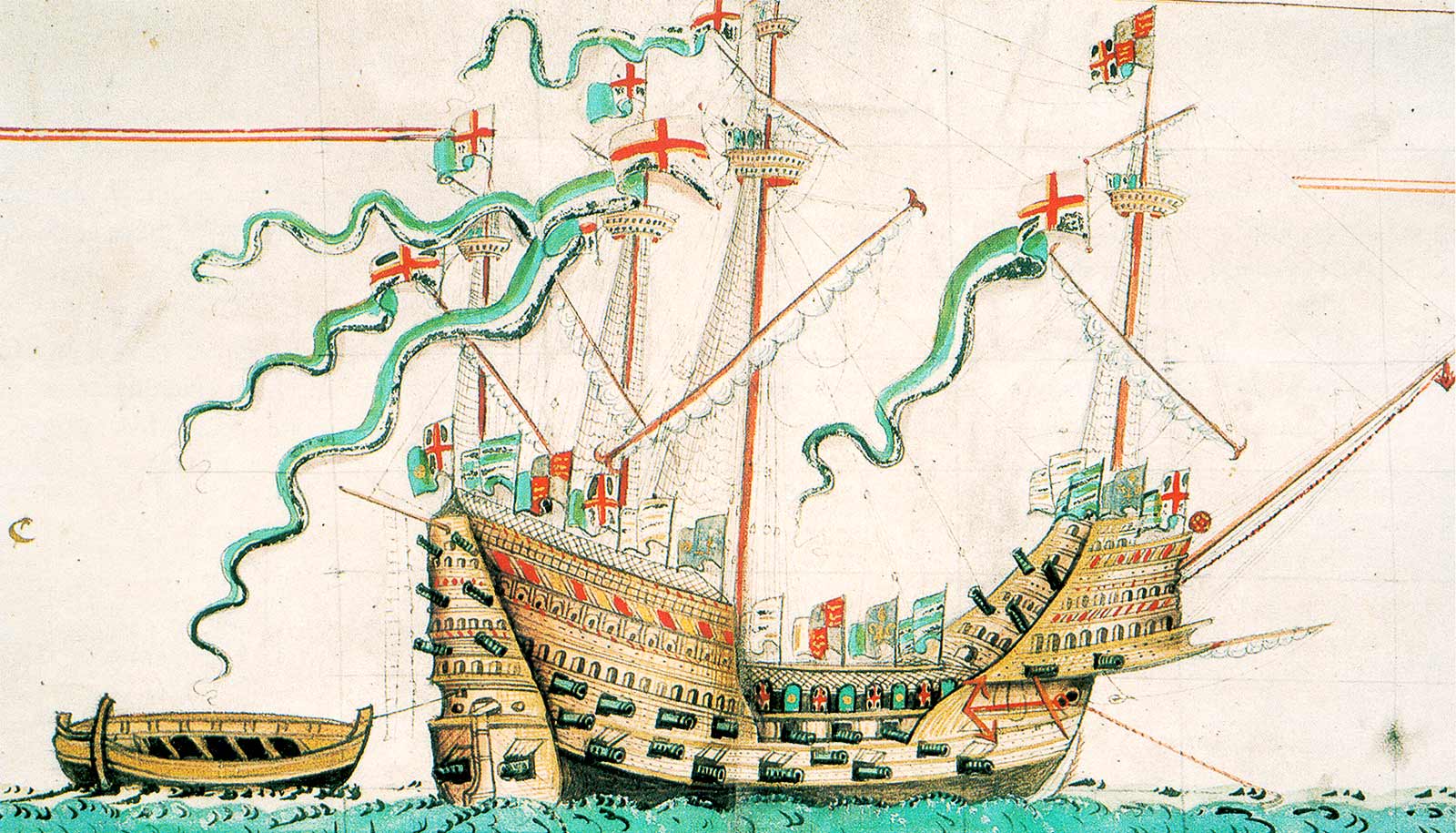
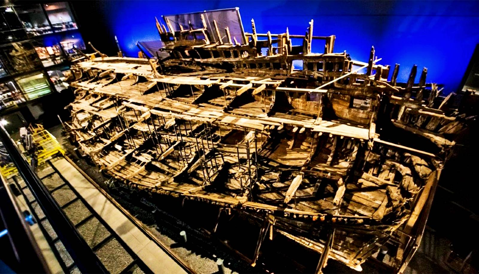
Though she ploughed the Atlantic and battled with her heavy cannons for 34 years—and laid buried beneath the turbulent English Channel for 437 more—bacteria and chemicals have begun eating away at her remnants, on display at the Mary Rose Museum in Portsmouth, England.

More than 500 years after its launch, the vessel remains a precious cultural treasure.
Among those involved in the project is Kirsten Marie Ørnsbjerg Jensen, associate professor at the University of Copenhagen, who leads the Nanostructure Group, where her research focuses on the internal structures of materials, right down to the nanolevel. Here, she scans materials using advanced X-ray technology, among other techniques. This puts her on home turf for this atypical research project.
“We have analyzed timber from the Mary Rose by way of a new technique that is somewhat comparable to how a hospital’s CT scanner works. The difference is that our scanning method combines CT scanning with what’s known as X-ray scattering,” Ørnsbjerg Jensen says.
“It lets us analyze the structure of materials at an atomic level, which makes it possible to find and map substances within the ship’s wood. It gives us information about what is contributing to the wood’s decay, so that it can be better preserved in the future.”
The Mary Rose sank during a naval battle with France in 1543 and was excavated in 1982, in what remains the world’s most expensive salvaging operation. At the time, 19,000 objects from the Tudor period were fished from the sea along with the shipwreck.
Today, the remains of the ship are on display at the Mary Rose Museum in Portsmouth. But after more than 400 years at the bottom of the English Channel, the wooden ship’s hull is vulnerable to decomposition. The threat includes deposits from metal parts on the ship and bacteria which trigger acid attacks on the timbers. The same threat applies to other cultural artifacts around the world that degrade after being found.
Thanks to the new technique, known as “X-ray computed tomography with pair distribution function analysis” (ctPDF), hope remains for the legendary vessel as well as other cultural artifacts and archaeological relics around the world.
The method helped the researchers identify a number of destructive substances that decompose wood when it resurfaces and is exposed to oxygen once again. These include zinc sulfides.
“To find a way to stop these decomposition processes, it is important to know what the sulfides are composed of and where they are. This technique allows us to do just that,” says Ørnsbjerg Jensen.
The Mary Rose Trust, which is the foundation behind the purpose-built museum for the old warship, is excited about the new X-ray technique.
“Being able to peer into the timbers of the Mary Rose and decode not only the wood’s structure, but also, the degree of degradation, how the wood was treated in the past, and the impact of the marine environment, provides fascinating insight.
“It presents us with a wealth of knowledge that we can use to understand how materials respond to specific treatments and environments, and to devise strategies to keep our cultural heritage intact for future generations,” says Eleanor Schofield, professor and deputy chief executive officer of the Mary Rose Trust.
The study appears in the journal Matter. Additional coauthors are from the University of Sheffield, Columbia University, the European Synchrotron Radiation Facility, the Mary Rose Trust, and the University of Copenhagen.
Source: University of Copenhagen