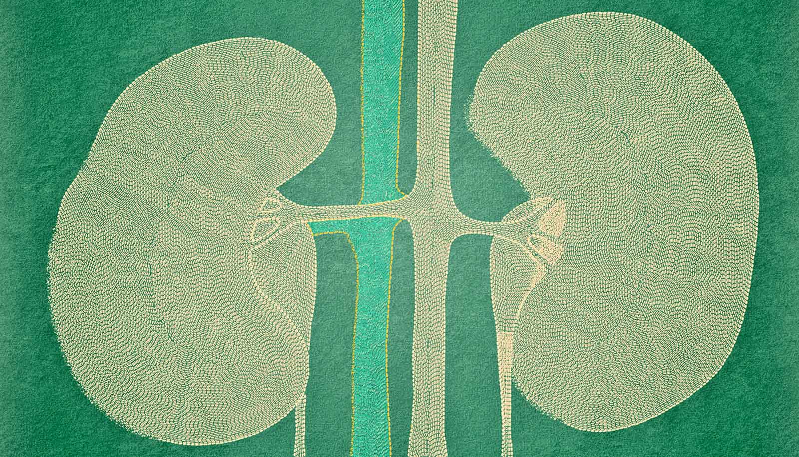
A new atlas that focuses on the kidney’s myriad cells could further understanding of kidney injury and disease.
Building on previous work showing 30 cell types in the kidney, the researchers revealed 51 cell types, some rare and novel, in the healthy kidney and 28 related cell types with features associated with injury or recovery.
They uncovered diverse cellular microcosms with rich genetic signatures along segments of the kidney associated with kidney failure in patients or recovery from injury. Altogether, they created a comprehensive 2D and 3D map of kidney cell organization and molecular identity in healthy and diseased kidneys.
“We don’t have great treatment options for patients with kidney disease,” says Sanjay Jain, a professor of medicine at Washington University in St. Louis and lead author of the study in Nature. “By mapping molecular signatures, we hope to predict which patients are at risk of progressing to kidney failure. This molecular knowledge will, one day, lead us to precise, customized treatments for our patients.”
Jain’s team worked with other members of the Kidney Precision Medicine Project (KPMP), Human BioMolecular Atlas Program (HuBMAP), Human Cell Atlas, and scientists from other institutions to complete single-cell and spatial tests on nearly 100 healthy and diseased human kidneys.
The Kidney Precision Medicine Project aims to improve kidney disease treatment. It is working to do so with the help of participants with kidney disease who are willing to undergo kidney biopsies solely to contribute to research. The study also received support from HuBMAP, an NIH Common Fund-sponsored program that aims to spatially define every cell in the healthy human body, and the Human Cell Atlas, an international research effort to gather information on at least 10 billion human cells.
The new research is one of nine studies, published simultaneously across the Nature Portfolio journals, introducing the first set of maps created by researchers at institutions supported by HuBMAP. Jain is also a co-senior author of another of the studies, published in Nature Communications, that maps healthy and injured cellular neighborhoods in areas where kidney stones form, and identifies biomarkers in the urine that are unique to patients with kidney stone disease.
Kidney functional mass slowly declines after age 35. Chronic illnesses such as diabetes and hypertension and accumulating kidney injury—through physiological aging, low oxygen, or changes in blood flow to the kidneys during surgery, reactions to medication, recreational drug use and even dehydration—can rapidly accelerate natural decline and cause kidney disease and, ultimately, kidney failure. Treatment options are limited to dialysis or difficult-to-come-by organ donation.
Using single-cell analyses, the researchers characterized the molecular features of healthy and diseased kidney cells in different kidney segments from patient kidney biopsy samples. They also performed spatial analyses to create 3D pictures of cells living in communities and communicating with their neighbors. A comprehensive view of such relationships may mitigate nonspecific therapeutic targeting of the whole kidney and entire cellular communities, and pave the way for better, more precise drugs with fewer side effects.
“We created a benchmark atlas that the community can use as a reference to get insights into how kidney disease develops,” says Jain, also a director of the Kidney Translational Research Center, which serves as a biorepository for collected kidney biopsies, some of which were used in the study as part of the Human BioMolecular Atlas Program.
“We looked at how kidney cells are organized, their molecular identities, and how they shift from healthy to diseased states. With this knowledge, we can start thinking about the drugs or small molecule targets that can prevent progression to disease or promote recovery from injury.”
Jain was surprised by the diversity of cell types, including interstitial, immune, and endothelial, uncovered by analyzing 300,000 human kidney cells.
“We found several altered cell states in many different kidney segments, reflecting the plasticity in cell phenotypes as cells transition through healthy, injury, and recovery states,” he says. The second version of the atlas, which is underway, involves analyzing more than 1 million kidney cells.
The collaboration also created several resources users can leverage to identify and understand their own research results. One such tool provides standardized nomenclature and benchmark identities of major, minor, and rare cells in the kidney. Researchers can now annotate their own data sets and help to promote consistent communication across research studies.
Additional coauthors are from Indiana University; the University of California, San Diego; the University of Michigan; and Washington University in St. Louis.
Funding came from the National Institutes of Health, the National Cancer Institute, the Institute of Clinical and Translational Sciences/Clinical and Translational Science, and the National Institute of Diabetes and Digestive and Kidney Diseases.