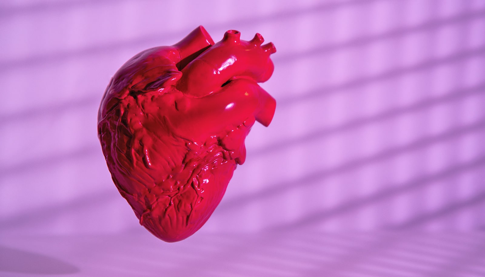
Heart defects—the most common type of birth defect—can result from mutations in the gene CHD4. Now, researchers know the key molecular details of what happens.
The CHD4 protein normally works in developing heart muscle cells to repress the production of muscle-filament proteins that are meant to operate in non-heart types of muscle cell. The failure of this repression leads to the development of abnormal, “hybrid” muscle cells that can’t pump blood as efficiently as normal heart cells.
“For patients with congenital heart defects linked to CHD4 mutations, this research helps explain why their hearts don’t work as well as normal, and suggests strategies for therapeutic intervention,” says senior author Frank Conlon, a professor in the biology and genetics departments at the University of North Carolina at Chapel Hill and a member of the UNC McAllister Heart Institute.
Researchers began by engineering mice whose developing embryos lack CHD4 just in their heart cells. The embryonic mice developed severe cardiac defects midway through gestation and none was born alive. These results confirmed the necessity for CHD4 in heart development.
CHD4, the protein encoded by the CHD4 gene, normally works as part of a multi-protein “machine” that helps regulate gene activity within the nuclei of cells, say researchers, who therefore conducted a set of experiments to measure and analyze the changes in developing heart-muscle cell gene activity when CHD4 is absent.
They found that the CHD4 protein normally binds directly to DNA in a way that represses the activity of genes that encode non-heart muscle proteins. These proteins help make up the springy fibers (myofibrils) that contract and relax when muscles work.
The team determined that when the CHD4 protein is absent, these other, non-cardiac muscle proteins are inappropriately produced in developing heart muscle cells. They become incorporated into the myofibrils in these cells, forming abnormal, hybrid myofibrils that lack the functional properties of the normal heart.
First author Caralynn M. Wilczewski, a graduate student in the Conlon laboratory, developed an advanced ultrasound technique and used it to record the activity of the tiny hearts developing in mice—organs which in mid-gestation are only about as large as the period at the end of this sentence.
“We observed that the hearts lacking CHD4 and having these abnormal cardiac myofibrils had severely reduced ventricular contractions, indicating a loss of the ability to pump blood normally,” Wilczewski says.
Blood vessels are key to building a strong heart
“These findings indicate that normal cardiac development in mice depends on the repression of non-cardiac myofiber proteins in heart muscle cells, to allow the formation of normal cardiac myofibers capable of sustaining normal heart contractions,” Conlon adds.
The findings, which appear in the Proceedings of the National Academy of Sciences, provide the first clear insight into the mechanism of CHD4-related cardiac defects. They also suggest the possibility that restoring the normal repression of non-cardiac myofiber proteins could prevent heart defects in cases where CHD4 is mutated.
How natural miscues cause heart defects in newborns
The researchers now plan to investigate the ways in which specific human CHD4 mutations lead to cardiac defects.
In addition, they plan to use the new ultrasound technology developed by Wilczewski in further research. “This technology has broad applications for testing models of congenital heart disease,” Conlon says.
Source: UNC-Chapel Hill