Scientists have made the first direct observation of a key step in the process bacteria use to rapidly develop antibiotic resistance and other traits.
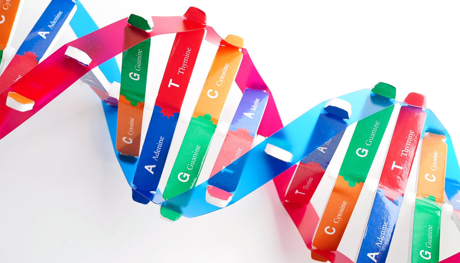
Researchers recorded the first images of bacterial appendages—over 10,000 times thinner than human hair—as they stretched out to catch DNA. The bacteria can then incorporate the DNA fragments into its own genome through a process called DNA uptake or “horizontal gene transfer.”
“Horizontal gene transfer is an important way that antibiotic resistance moves between bacterial species, but the process has never been observed before, since the structures involved are so incredibly small,” says senior author Ankur Dalia, an assistant professor in the College of Arts and Sciences’ biology department at Indiana University-Bloomington.
https://giphy.com/gifs/cmzl6Eey3LMkecrZHO
“It’s important to understand this process, since the more we understand about how bacteria share DNA, the better our chances are of thwarting it,” he says.
Antibiotic-resistant bacteria affects nearly 1 million people each year, according to the World Health Organization. WHO has found evidence of these strains in nearly 490,000 people with tuberculous and 500,000 people with other infectious diseases.
The bacterium the researchers used in the study was Vibrio cholerae, the microbe that causes cholera. The structures bacteria use to catch DNA in the environment are extremely thin, hair-like appendages called pili.
Although scientists were aware that pili play a role in DNA uptake, direct evidence demonstrating how they work was lacking until this study, Dalia says. In order to observe pili in action, the scientists “painted” both the pili and DNA fragments with special glowing dyes.
Understanding bacteria ‘switch’ could lead to new antibiotics
The new study uses these dyes to reveal that pili act like microscopic “harpooners” that cast their line through pores in the cell’s wall to “spear” a stray piece of DNA at the very tip. The pili then “reel” the DNA into the bacterial cell through the same pore.
The pore is so small that the DNA would need to fold in half to fit through the opening in the cell, Dalia says.
“It’s like threading a needle,” says Courtney Ellison, a PhD student and first author of the study, which appears in Nature Microbiology.
“The size of the hole in the outer membrane is almost the exact width of a DNA helix bent in half, which is likely what is coming across. If there weren’t a pilus to guide it, the chance the DNA would hit the pore at just the right angle to pass into the cell is basically zero.”
The team wants to next study exactly how pili “hook” onto the DNA at just the right spot, especially since the protein involved in the process appears to interact with DNA in an entirely new way, Dalia says. His team also looks forward to applying their pili labeling method to study other functions played by these diverse bacterial structures.
Is ‘antivirulence’ the answer to failing antibiotics?
Distinguished professor Yves Brun is also a lead author of the paper. Other coauthors are from Indiana University and City University New York Brooklyn. The National Institutes of Health and the National Science Foundation supported the work.
Source: Indiana University