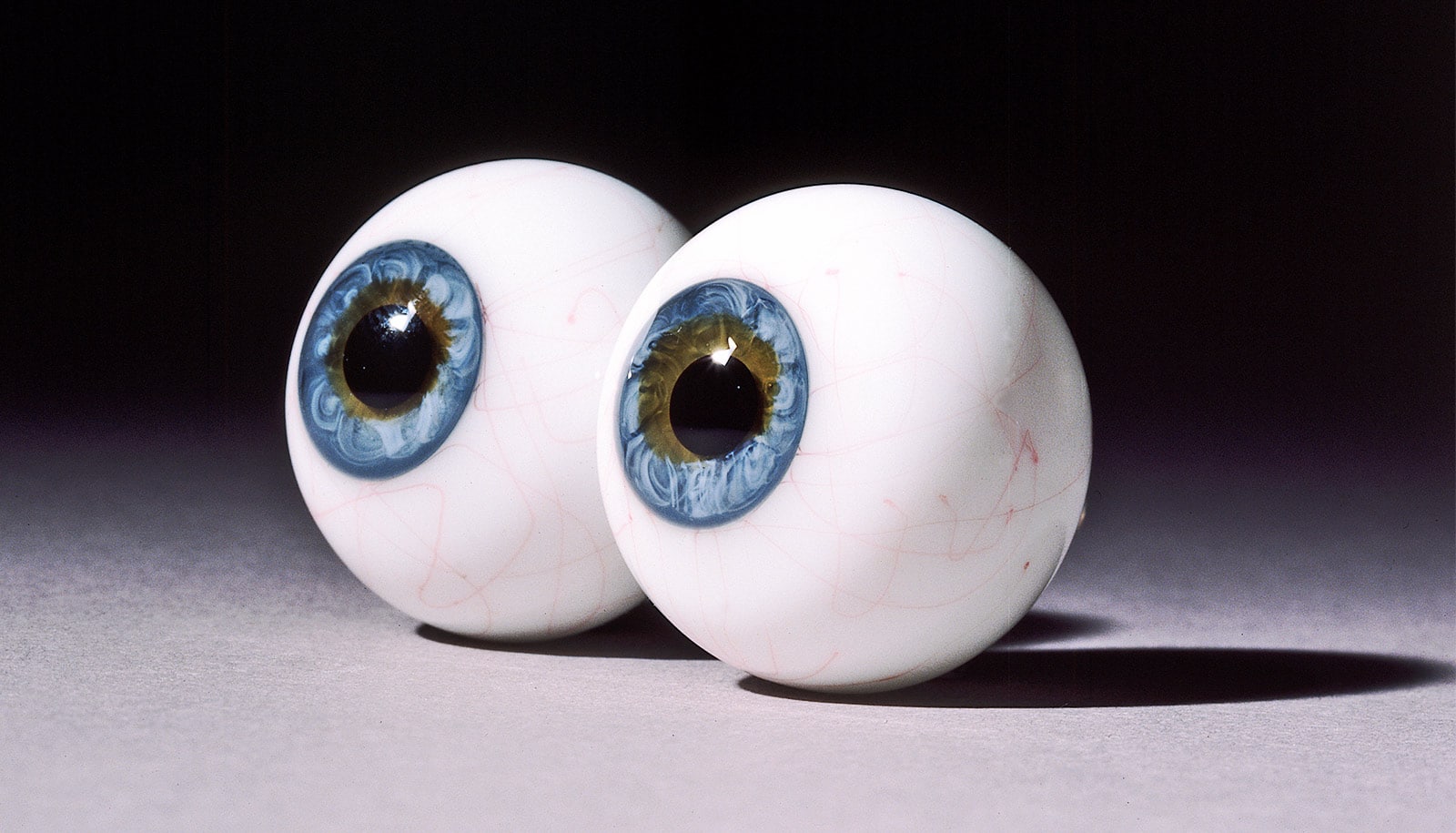
Researchers have identified elusive cells in the eye that could explain how humans see red, green, blue, and yellow.
Scientists have long wondered how the eye’s three cone photoreceptor types work together to allow humans to perceive color.
In the new study in the Journal of Neuroscience, researchers at the University of Rochester used adaptive optics to identify rare retinal ganglion cells (RGCs) that could help fill in the gaps in existing theories of color perception.
The retina has three types of cones to detect color that are sensitive to either short, medium, or long wavelengths of light. Retinal ganglion cells transmit input from these cones to the central nervous system.
In the 1980s, David Williams, a professor of medical optics, helped map the “cardinal directions” that explain color detection. However, there are differences in the way the eye detects color and how color appears to humans. Scientists suspected that while most RGCs follow the cardinal directions, they may work in tandem with small numbers of noncardinal RGCs to create more complex perceptions.
The new research suggests some of these elusive noncardinal RGCs in the fovea that could explain how humans see color.
“We don’t really know anything for certain yet about these cells other than that they exist,” says Sara Patterson, a postdoctoral researcher at the University of Rochester Center for Visual Science who led the study. “There’s so much more that we have to learn about how their response properties operate, but they’re a compelling option as a missing link in how our retina processes color.”
The team leveraged adaptive optics, which uses a deformable mirror to overcome light distortion and was first developed by astronomers to reduce image blur in ground-based telescopes. In the 1990s, Williams and his colleagues began applying adaptive optics to study the human eye. They created a camera that compensated for distortions caused by the eye’s natural aberrations, producing a clear image of individual photoreceptor cells.
“The optics of the eye’s lens are imperfect and really reduce the amount of resolution you can get with an ophthalmoscope,” says Patterson. “Adaptive optics detects and corrects for these aberrations and gives us a crystal-clear look into the eye. This gives us unprecedented access to the retinal ganglion cells, which are the sole source of visual information to the brain.”
Patterson says improving our understanding of the retina’s complex processes could ultimately help lead to better methods for restoring vision for people who have lost it.
“Humans have more than 20 ganglion cells and our models of human vision are only based on three,” says Patterson. “There’s so much going on in the retina that we don’t know about. This is one of the rare areas where engineering has totally outpaced visual basic science. People are out there with retinal prosthetics in their eyes right now, but if we knew what all those cells do, we could actually have retinal prosthetics drive ganglion cells in accordance with their actual functional roles.”
Support for the work came from the National Institutes of Health, Air Force Office of Scientific Research, and Research to Prevent Blindness.
Source: University of Rochester