Using microscopy and mathematics, researchers have discovered the invisible pattern that growing neurons follow during brain formation.
The technique could one day allow bioengineers to coax stem cells to grow into replacement artificial tissues and organs.
Life is rife with patterns. It’s common for living things to create a repeating series of similar features as they grow: think of feathers that vary slightly in length on a bird’s wing or shorter and longer petals on a rose. It turns out the brain is no different.
The new study in Nature Physics builds on the fact that the brain contains many different types of neurons and that it takes several types working in concert to perform any tasks. The researchers wanted to uncover the invisible growth patterns that enable the right kinds of neurons to arrange themselves into the right positions to build a brain.
“How do cells with complementary functions arrange themselves to construct a functioning tissue?” says coauthor Bo Wang, an assistant professor of bioengineering at Stanford University.
“We chose to answer that question by studying a brain because it had been commonly assumed that the brain was too complex to have a simple patterning rule. We surprised ourselves when we discovered there was, in fact, such a rule.”
The brain they chose to examine belonged to a planarian, a millimeter-long flatworm that can regrow a new head every time after amputation.

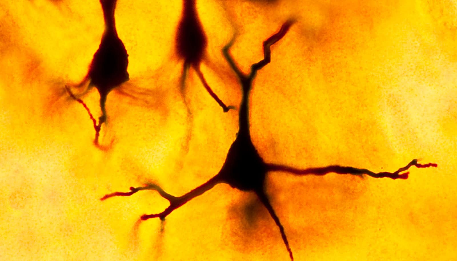
First, Wang and Margarita Khariton, a graduate student in his lab, used fluorescent stains to mark different types of neurons in the flatworm. They then used high-resolution microscopes to capture images of the whole brain—glowing neurons and all—and analyzed the patterns to see if they could extract from them the mathematical rules guiding their construction.
They found that each neuron is surrounded by roughly a dozen neighbors similar to itself, but that other kinds of neurons are interspersed among them. This unique arrangement means that no single neuron sits flush against its twin, while still allowing different types of complementary neurons to be close enough to work together to complete tasks.
The researchers found that this pattern repeats over and over across the entire flatworm brain to form a continuous neural network.
Coauthors Jian Qin, an assistant professor of chemical engineering, and postdoctoral scholar Xian Kong developed a computational model to show that this complex network of functional neighborhoods stems from the tendency of neurons to pack together as closely as possible without being too close to other neurons of the same type.
While neuroscientists might someday adapt this methodology to study neuronal patterning in the human brain, the researchers believe the technique could more usefully apply to the emerging field of tissue engineering.
The basic idea is simple: tissue engineers hope to induce stem cells, the powerful, general-purpose cells from which all cell types derive, to grow into the various specialized cells that form a liver, kidney, or heart. But scientists will need to arrange those diverse cells into the right patterns if they want the heart to beat.
“The question of how organisms grow into forms that carry out useful functions has fascinated scientists for centuries,” Wang says. “In our technological era, we are not limited to understanding these growth patterns at the cellular level but can also find ways to implement these rules for bioengineering applications.”
The Burroughs Wellcome Fund funded the work.
Source: Stanford University