Researchers have identified important molecules that can help identify specific glial cells integral to synapses in the brain.
While the brain is composed of two types of cells, glia tend to receive far less attention for their importance in brain function and disease than the more celebrated neurons.
Scientists have known for more than a century, however, that special types of glial cells are integral components of neuromuscular junctions or synapses—points of contact between neurons and muscles that permit the brain to control movement.
Despite the significance of glial cells for the proper formation, maintenance, and repair of synapses throughout the nervous system, the inability to distinguish specific glial cells at synapses from the diverse overall population of glial cells has been a major challenge in promoting and restoring the normal function of the nervous system following injuries, diseases and in old age.
The new research could change that.
“This discovery will serve as a springboard to addressing fundamental questions and developing assays to speed the discovery of therapeutics intended to preserve and restore the normal function of neuronal circuits,” says Gregorio Valdez, an associate professor of molecular biology, cell biology, and biochemistry at Brown University who is affiliated with the new Center for Translational Neuroscience, which the Carney Institute for Brain Science at Brown and the Brown Institute for Translational Science established.
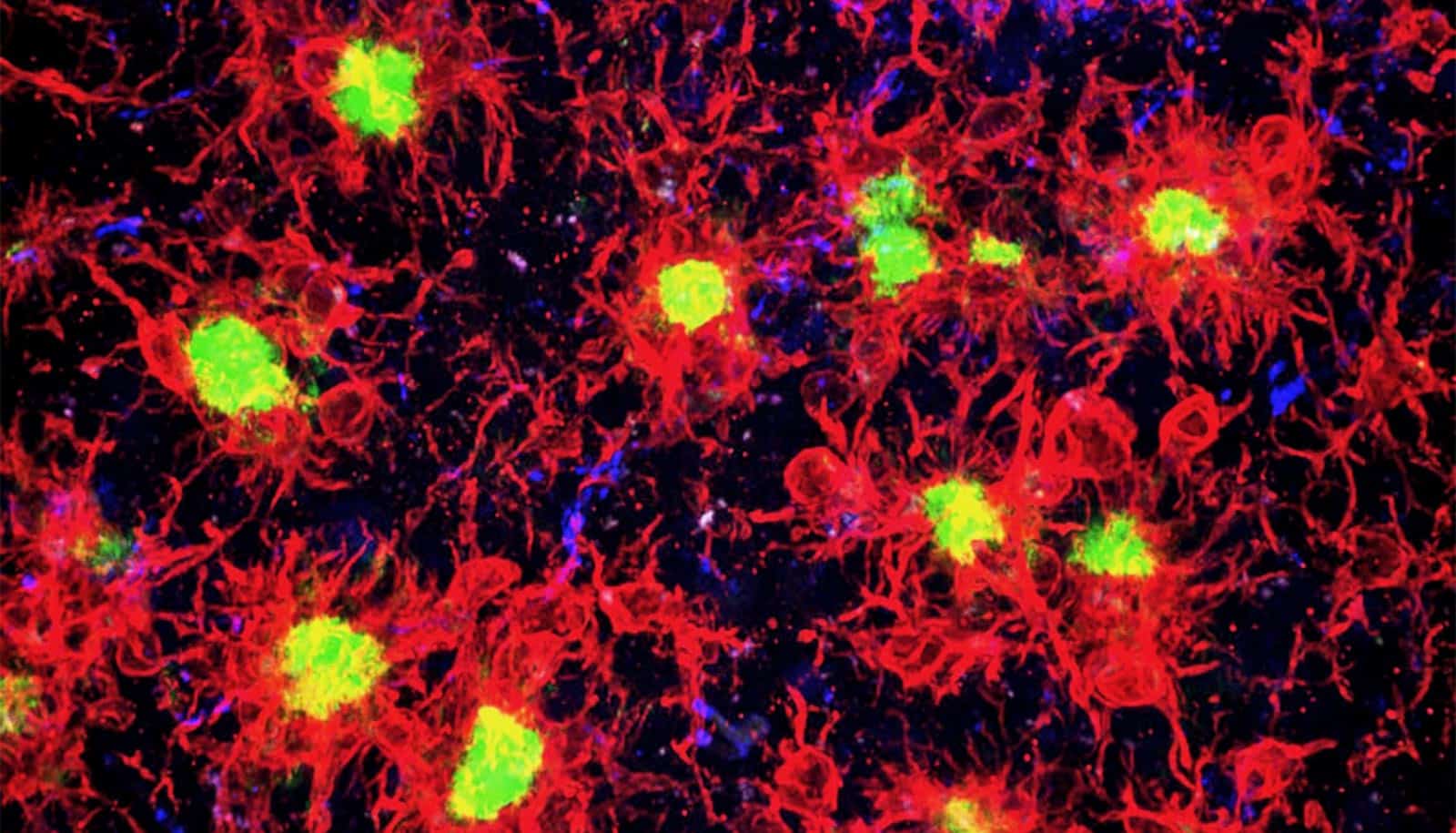
The study reveals that an important subtype of glia, known as Schwann cells and located at neuromuscular synapses, are the only cells in muscles expressing two specific molecules.
These molecular markers provide a highly specific glial “barcode,” Valdez says, that identifies the vital cell subtype.
“What this means is that we can finally figure out how all three cellular constituents of the synapse—neurons, muscle, and glia—talk to each other,” Valdez says.
“We now have a unique and important tool for identifying this critical component of the synapse. This is essential for knowing when and where to target to ensure synapses function appropriately.”
Valdez says the novel barcode tool will pave the way for future studies, including on neuromuscular diseases such as amyotrophic lateral sclerosis (ALS) and spinal muscular atrophy (SMA). Scientists can use the molecular markers to probe the role of synaptic glia in neuromuscular synapse repair following injury, degeneration during normal aging, and the progression of neuromuscular diseases.
He also anticipates that a similar approach will reveal the synaptic glial cells located at synapses between pairs of neurons in the brain.
“While our primary focus was the neuromuscular synapse, we also gathered initial evidence indicating that synaptic glia cells in the brain can be labeled and targeted using the same approach,” he says.
“If true, this discovery could be of immense consequence for treating a myriad of brain conditions, including those involving cognitive decline due to normal aging and Alzheimer’s Disease.”
The research appears in the journal eLife. Additional coauthors are from Brown and Virginia Tech.
Funding for the study came from the National Institute on Aging and the National Institute of Neurological Disorders and Stroke.
Source: Brown University