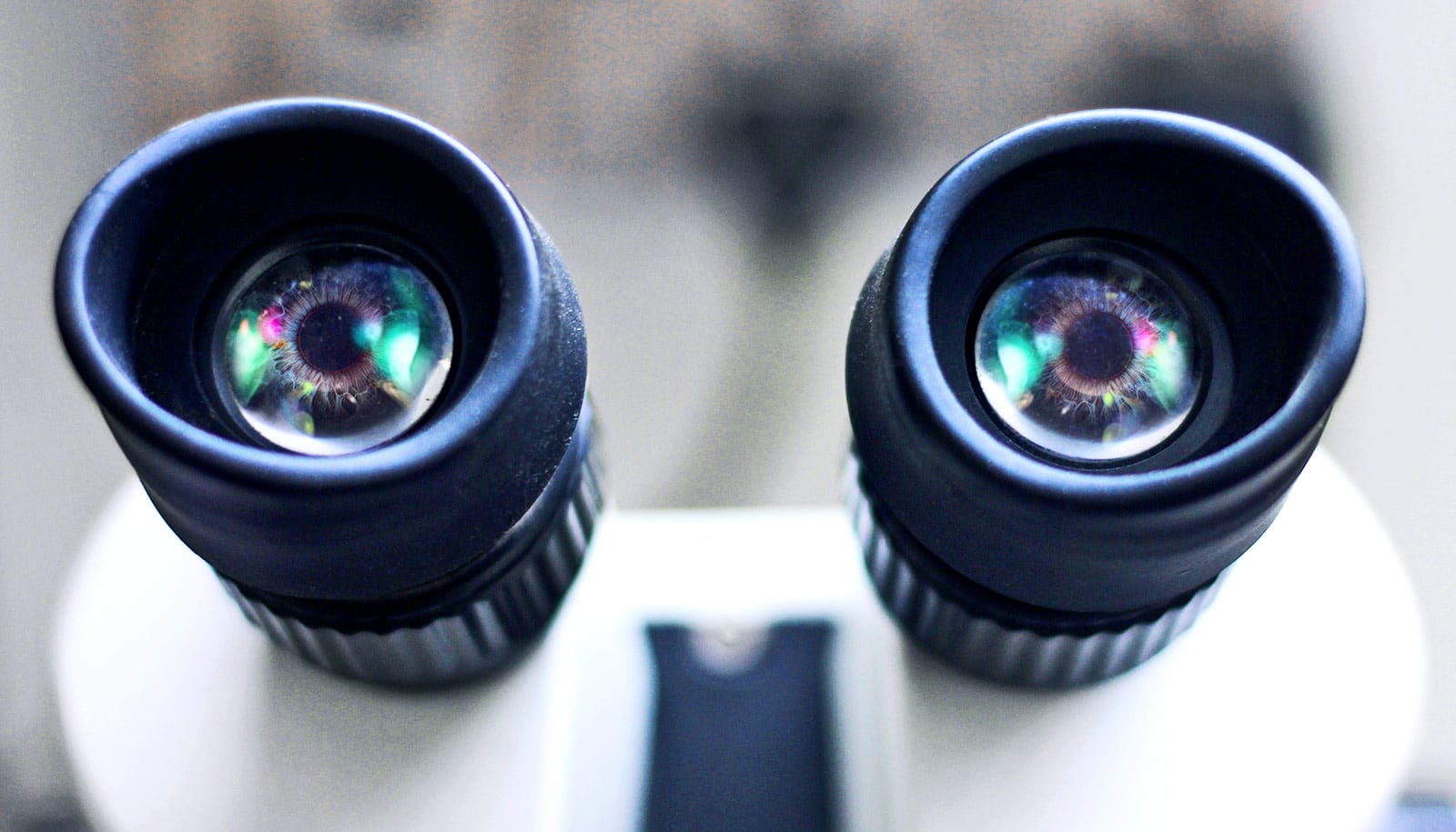
A specialized microscope is bringing scientists one step closer to understanding cell behavior, according to a new study.
Previously, in order to study cell membranes, researchers would often have to freeze samples—but the proteins within the samples wouldn’t behave like they would in a normal biological environment.
Now, using an atomic force microscope, researchers can observe individual proteins in an unfrozen sample—acting in a normal biological environment. This new observation tool could help scientists better predict how cells will behave when new components are introduced.
Real-time 3D look
“What’s missing right now in cell biology is the ability to predict cell behavior,” says Gavin King, associate professor of physics and astronomy at the University of Missouri.
“We don’t know all of the details yet on a number of biological processes. For example, when a drug is introduced to a cell, it must pass through the membrane, which may create a reaction. The more knowledge we have about that reaction, the better we will be able to create drugs that can target a specific area and, possibly, result in fewer side effects,” King says.
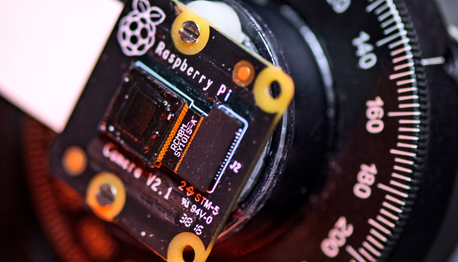
The atomic force microscope is capable of tracing the three-dimensional shape of an individual protein in biological conditions (in fluid at room temperature). It’s comprised of a robotic arm with a tiny needle attached on one end.
Researchers position the arm precisely on the sample they wish to analyze. Then, by very gently tapping the needle multiple times into the specimen in various points, a real-time, three-dimensional image of a protein is developed.
Molecular ‘movie’
For the current study, which appears in Science Advances, researchers focused on imaging the consequences of a chemical reaction occurring within one particular protein from E.coli that is responsible for transporting other proteins across the cell membrane.
They picked E.coli because of the simplicity of its cells. While researchers couldn’t control the precise moment the reaction occurred, the force microscope’s tapping motion allowed researchers to watch in real time how that protein changed its shape in response to the release of chemical energy. These conformational changes are directly related to the protein’s biological function.
“We can keep our eyes on just one protein, add various components, and then watch what happens,” King says. “It is like making a movie of a single molecule doing its biological work. We are really in the early days of understanding the mechanical details of how cells work, but as these tools become increasingly more precise they could provide us with essential information in the future.”
The National Science Foundation and a Burroughs Welcome Fund Career Award funded the work. The content is solely the responsibility of the authors and does not necessarily represent the official views of the funding agencies.
Source: University of Missouri


