A rare group of neurons can reconnect broken spinal circuits and trigger leg muscle activity after spinal cord injury, according to new research.
The discovery could help refine future stem-cell therapies for paralysis.
The findings show that certain neurons derived from transplanted neural stem cells can integrate into the spinal cord’s motor networks and relay signals to muscles responsible for walking.
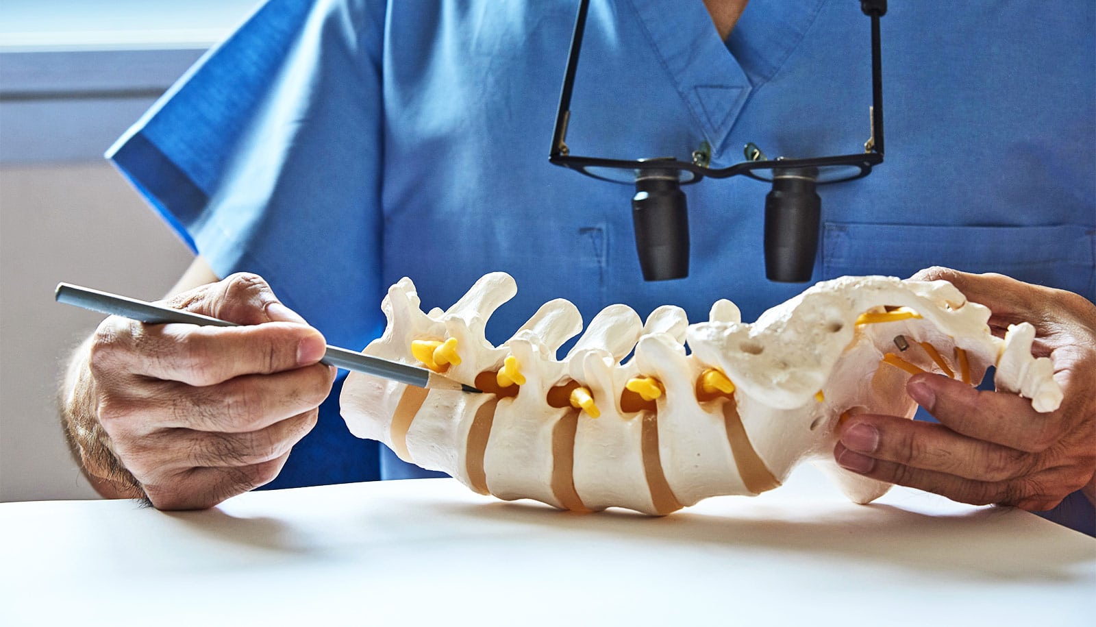
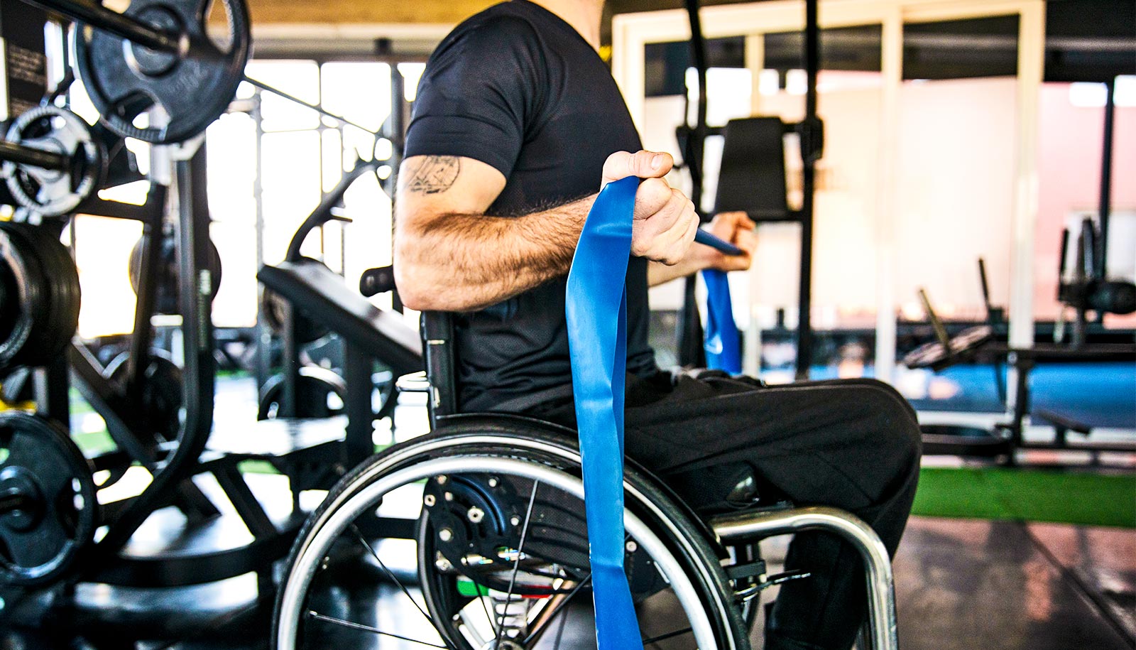
Spinal cord injuries occur when trauma damages the bundle of nerves that carries signals between the brain and the rest of the body, cutting off communication with muscles and organs below the injury. The result is often permanent paralysis along with a cascade of other medical implications.
Despite decades of research, there are currently no FDA-approved therapies that restore lost neurological function after spinal cord injury, leaving hundreds of thousands of people in the United States with lifelong disability.
For years, scientists have explored transplanting neural stem cells into injured spinal cords in hopes that the new neurons could replace damaged ones and rebuild lost connections. What has remained unclear is which cells within those grafts connect to the spinal cord’s walking circuits.
By tracking how transplanted neurons connect with spinal motor circuits and identifying the interneuron subtypes capable of activating leg muscles, this study begins to pinpoint the specific neuron types capable of rebuilding those pathways.
“Imagine an electrical circuit with a battery on one end and a light bulb on the other,” says Jennifer Dulin, assistant professor of biology at Texas A&M University and the study’s senior author.
“If the wires between them are disconnected, the light bulb won’t turn on. A spinal cord injury breaks that circuit. What we’re trying to do is place new cells into the middle so they can reconnect the pathway and allow signals to flow again.”
In the new study, scientists transplanted neural progenitor cells into injured spinal cords in animal models and examined how the transplanted cells connected to surrounding nerve networks. The research examined how graft-derived neurons connect to spinal motor circuits that control the hind limbs.
When a small subset of these transplanted neurons was experimentally activated, the animals’ leg muscles responded—evidence that the grafted cells had become part of the spinal cord’s motor circuitry.
The team also discovered that these crucial interneurons were relatively rare in the transplanted cell population. In the study, leg muscle responses were observed in roughly 20% to 30% of animals. Dulin says even that level is significant.
“This is meaningful because it shows the potential to recreate these walking neural circuits is there,” she says. “The next step is understanding why some animals respond to the treatment and others don’t.”
The findings could help guide the next generation of regenerative therapies by revealing which specific neurons need to be enriched in transplanted cell populations. The research also highlights another factor that may shape recovery: rehabilitation.
Newly transplanted neurons are immature and must adapt to the spinal cord’s environment, Dulin says, a process that depends on activity.
“We’re essentially putting newborn neurons into the spinal cord, and they don’t have any experience yet,” she says.
“Just like babies learn by interacting with their environment and practicing movements, these transplanted neurons need activity to learn how to function within the circuit.”
Pairing targeted cell therapies with activity-based rehabilitation could therefore be essential for helping transplanted neurons integrate effectively into the body’s existing motor networks.
“This kind of basic biology research is critically needed in order to develop new therapies,” Dulin says.
“For decades in the field of spinal cord injury we’ve just been testing treatments without really understanding how they work. We’re entering a new era where we have amazing tools to really study the effects of a treatment on an individual cellular level. These kinds of studies are critical to paving the way for effective human treatments.”
The research appears in Nature Communications.
Source: Texas A&M University