Microbubbles measured in microns—millionths of a meter—can form in cerebral spinal fluid inside the skull during traumatic brain injuries, according to new research.
The “formation and dramatic collapse” of these microbubbles could be responsible for some of the damage in a brain injury, the researchers report.
Bubble damage may sound trivial. But bubble collapse—a process known as cavitation— and the resulting shock waves can damage the steel foundations of boat propellers.
The researchers report that prior studies indicate the expansion and collapse of microbubbles creates forces of 0.1 to 20 megapascals, or 14.5 to 2,900 pounds per square inch.
“…[S]o it is alarming to realize the damage that cavitation inflicts on vulnerable brain tissue,” the researchers write.
TBI and microbubbles
To test and characterize the impact of cavitation inside the skull, the researchers simulated a brain by creating a 3D cell culture platform for astrocytic cells (star-shaped cells in the brain and spinal cord that are active in supporting, maintaining and repairing the central nervous system). They submerged the cell culture platform in a small tank and created microbubbles around 60 millionths of a meter in size. Some of the microbubbles adhered to the cell-laden microfiber scaffold.
Researchers turned on an ultrasonic device in the tank, collapsing the microbubbles and creating cavitation. (They also used the ultrasonic device on a control group of cells that were not exposed to cavitation.)
The researchers looked for two kinds of effects:
First, they used an inverted microscope to record any morphological changes to the cells. Second, they worked with colleagues in Iowa State’s College of Veterinary Medicine to assess whether there were genetic changes in the cells.
The researchers found cavitation caused the cells to shrink and roughened their surfaces. The cells appeared to elongate and grow when images were taken 22 and 48 hours after cavitation. Even so, the researchers found cell growth after 48 hours—in terms of surface area—was about half as much as the control cells.
The researchers also found the cells damaged by cavitation had elevated expression of genes such as TNF-α and IL-6, which are associated with inflammatory conditions such as infection with the SARS-CoV-2 virus and neurological disorders such as Parkinson’s and Alzheimer’s diseases.
“Taken together, these results confirm that surrounding cavitation is detrimental to astrocytic function,” the researchers write.
Ways to limit the damage
Hashemi says while doctors look for treatments for the brain damage cavitation causes, she says engineers can work to identify the places in the brain where cavitation is most likely to occur.
“A location map of cavitation occurrence can be directly used to design a helmet that dampens force and reduces the possibility of cavitation,” the researchers write.
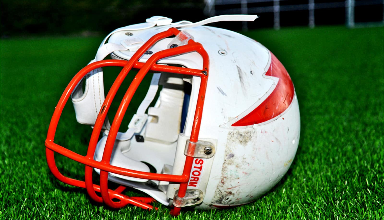
And while this study focusses on military helmets, Hashemi says the same ideas could be applied to helmets for football and other sports.
“This research is looking at battlefield applications, but in football there are similar impacts and shock waves,” Hashemi says. “Players do get mild forms of traumatic brain injury. Players might not realize it, but the effects of cavitation injuries would be there.”
Alex Wrede, Hashemi’s former graduate student who’s now working as a dynamic systems modeling engineer for John Deere in Dubuque, says the project has taught him there’s great need for more research and development.
“The people who have served our country and come back with injuries are really relying on research to find answers,” he says. “Answers could improve quality of life for our veterans and everybody unfortunate enough to go through traumatic brain injury.”
The findings appear in Global Challenges. Additional coauthors are from Iowa State and Clinton, Illinois High School. Support for the research came from the Office of Naval Research.
Source: Iowa State University