Analysis of sawfish fossils may help clarify the evolution of teeth.
Did they evolve from body scales that migrated into the mouths of ancient vertebrates and became adapted for eating—an idea known as the “outside-in” hypothesis?
Or did they evolve independent of scales, originating deep within the oral cavity and ultimately mounting onto the jaws—known as the “inside-out” hypothesis? A new study provides evidence for the “outside-in” hypothesis.
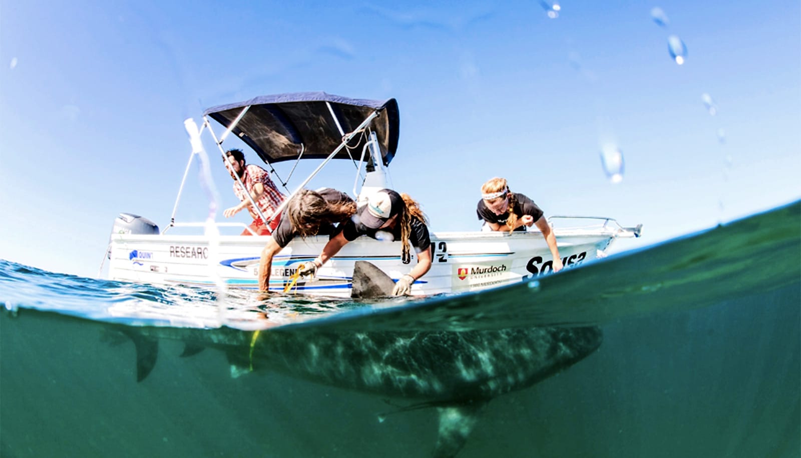
According to vertebrate paleontologist Todd Cook, associate professor of biology at Penn State Behrend, the team didn’t set out to contribute to the debate about the origins of teeth. Instead, he and his colleagues were studying the tissue structure of rostral denticles, which are the jagged spikes that run along the elongated snouts of sawsharks and sawfishes. The animals use them in foraging and self-defense.
Cook, lead author of the study in the Journal of Anatomy, notes that sawfishes belong to the same group as skates and rays and are closely related to sharks.
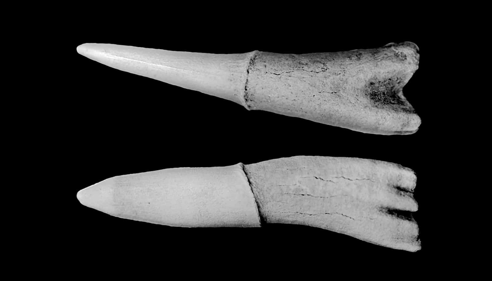
The team examined the fossilized rostral denticles of Ischyrhiza mira, a species belonging to an extinct group of sawfishes that lived in North American waters during the late Cretaceous period, around 100 to 65 million years ago. The samples had previously been recovered from a rock formation in New Jersey.
“Rostral denticles are believed to be modified scales because of their location on the elongated snout and they have an external morphology and developmental pattern that’s similar to scales,” says Cook, who explains that, just like with scales found elsewhere on the body, for a new rostral denticle to form, an old one must first fall off and make a space available.
“Yet, very little was known about the organization of the tissues that make up rostral denticles, particularly the hard outermost layer known as enameloid.

“Given that rostral denticles are likely specialized body scales, we hypothesized that the enameloid of rostral denticles would exhibit a similar structure to the enameloid of body scales, which have simple microcrystal organization.”
To examine the internal microstructure of the fossil rostral denticles, the researchers hand sectioned the samples, both transversely—across the width—and longitudinally—across the length. Next, they used a scanning electron microscope to study the histology—or microscopic anatomy—of the rostral denticles.
“Surprisingly, Ischyrhiza mira’s rostral denticle enameloid was anything but simple; it was considerably more complex than the enameloid of body scales,” says Cook. “In fact, the overall organization of the enameloid in this ancient sawfish resembled that of modern shark tooth enameloid, which has been well-characterized.”
Specifically, he notes that both Ischyrhiza mira rostral denticles and modern shark teeth have an enameloid covering that largely consists of fluorapatite microcrystals packed together into distinct bundles. Towards the outer region of the enameloid, these bundles run parallel to the surface of the tooth and are called the “parallel bundled enameloid.” Deeper, the bundles become randomly arranged, a region known as the “tangled bundled enameloid.” Finally, passing through these layers is the “radial bundled enameloid,” which is composed of packed microcrystals oriented perpendicular to the tooth surface.
In terms of function, Cook explains that having bundles of microcrystals arranged in various orientations enables shark teeth to resist the mechanical stresses associated with feeding. Similarly, he notes, “It is likely that the bundled microcrystal arrangement of the enameloid of Ischyrhiza mira’s rostral denticles also served as a way to withstand mechanical forces.”
However, the most surprising and consequential outcome of this study is that it makes an important contribution to the long-standing debate regarding the origin of teeth, says Cook. Specifically, he explains, “This finding provides direct evidence supporting the ‘outside-in’ hypothesis, as it shows that scales have the capacity to evolve a complex tooth-like enameloid outside of the mouth. It is more parsimonious to suggest that scales produced a similar bundled microstructure in teeth and rostral denticles than to conclude that both these structures evolved a similar enameloid independently.”
A Penn State Behrend Undergraduate Research Grant supported this research.
Source: Penn State