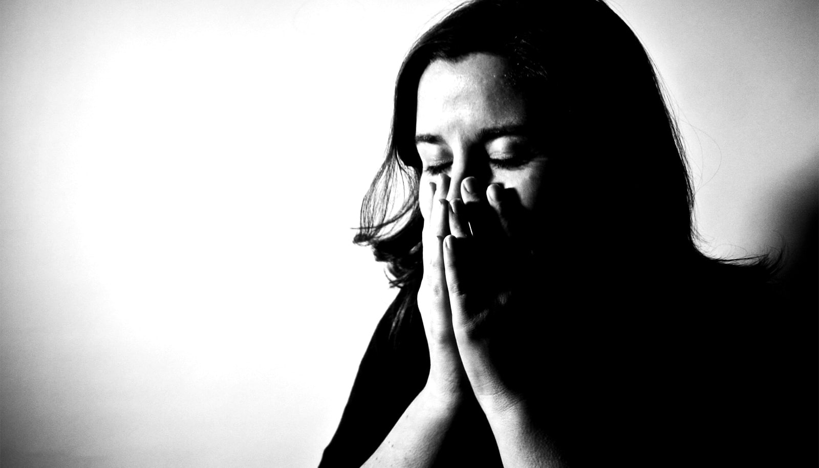
Researchers have found a home in the brain for the feeling of stress.
The new research may help people deal with the debilitating sense of fear and anxiety that stress can evoke, researchers report.
Brain scans of people exposed to highly stressful and troubling images—such as a snarling dog, mutilated faces, or filthy toilets—reveal a network of neural connections emanating throughout the brain from the hippocampus, an area of the brain that helps regulate motivation, emotion, and memory.

The brain networks that support the physiological response to stress have been well studied in animals. Activation of brain areas such as the hypothalamus triggers production of steroid hormones called glucocorticoids in the face of stress and threats. But the source of the subjective experience of stress experienced by people during the COVID-19 pandemic, for instance, has been more difficult to pinpoint.
“We can’t ask rats how they are feeling,” says lead author Elizabeth Goldfarb, associate research scientist at the Yale University Stress Center.
The researchers conducted a series of fMRI scans of subjects who they asked to quantify their stress levels when presented with troubling images.
The study reveals that neural connections emanating from the hippocampus when viewing these images reached not only areas of the brain associated with physiological stress responses, but also the dorsal lateral frontal cortex, an area of the brain involved in higher cognitive functions and regulation of emotions.
The researchers found that when neural connections between the hippocampus and frontal cortex were stronger, subjects reported feeling less stressed by the troublesome images.
Conversely, subjects reported feeling more stressed when the neural network between the hippocampus and hypothalamus was more active.
The authors note there is also evidence from other studies that those suffering from mental health disorders such as anxiety may have difficulty receiving calming feedback from the frontal cortex in times of stress.
“These findings may help us tailor therapeutic intervention to multiple targets, such as increasing the strength of the connections from the hippocampus to the frontal cortex or decreasing the signaling to the physiological stress centers,” says Rajita Sinha, a professor of psychiatry and professor in Yale’s Child Study Center and neuroscience department.
All study subjects were healthy, she says, and in some cases their responses during the experiment seemed to be adaptive—in other words, the network connections with the frontal cortex became stronger as the subjects were exposed to the stressful images. Sinha and Goldfarb speculated that these subjects might be accessing memories that help moderate their response to stressful images.
“Similar to recent findings that remembering positive experiences can lower the body’s stress response, our work suggests that memory-related brain networks can be harnessed to create a more resilient emotional response to stress,” Goldfarb says.
The research appears in Nature Communications.
Source: Yale University