To rehearse for complex surgery on the spinal cords of fetuses, surgeons are practicing on kickballs and chicken breasts.
These items, combined with high-tech 3D printing technology, are providing surgical teams with the practice they need to perform a complex but minimally invasive new surgical repair of a particular form of spina bifida.
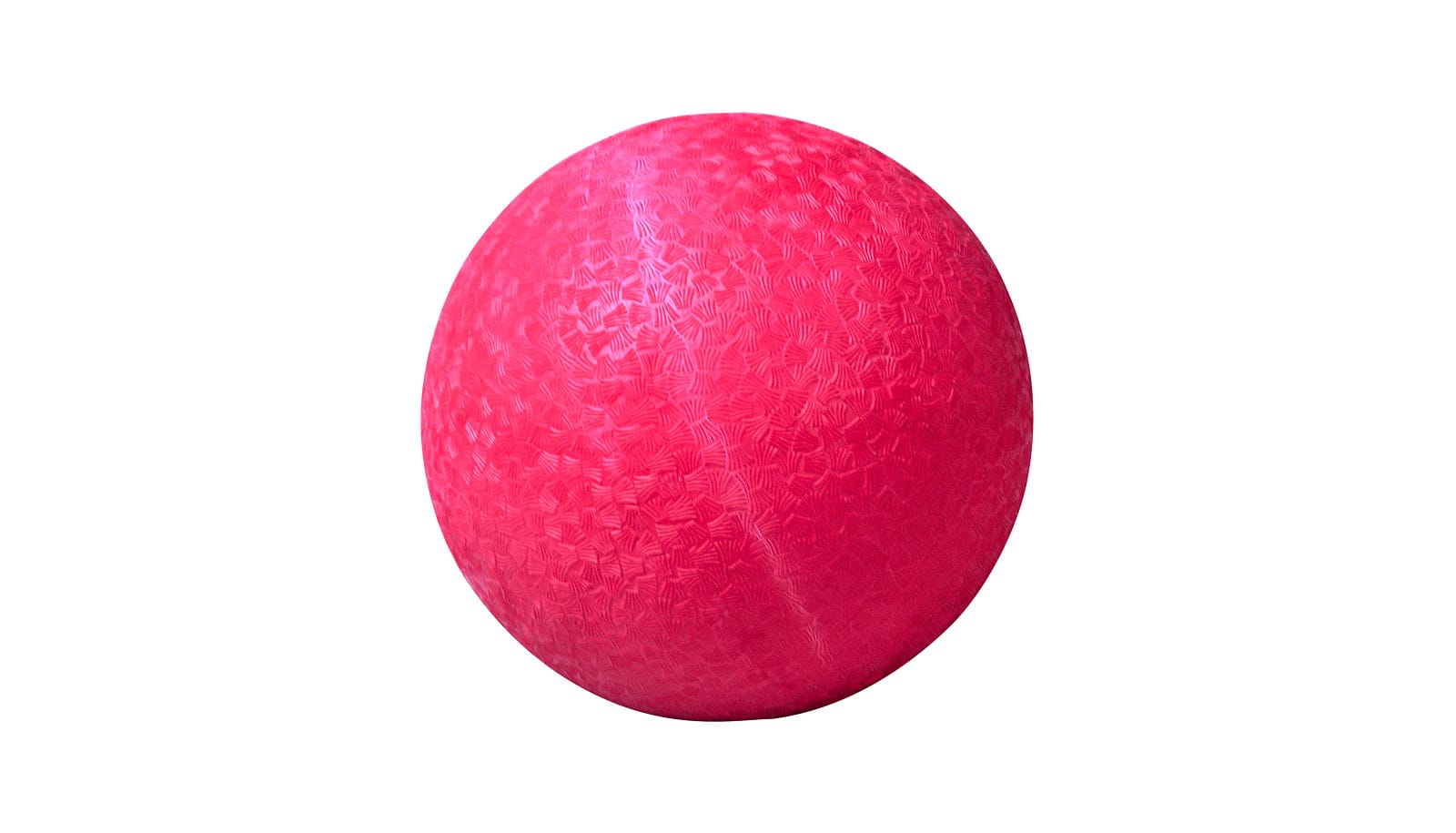
“A kickball is about the size of a uterus at that time in pregnancy.”
Spina bifida is the failure of the spinal column to close normally during early fetal development. It occurs in about three to four of every 10,000 pregnancies. It can result in permanent nerve damage if left untreated.
The new procedure, called fetoscopic myelomeningocele repair, involves a maternal fetal medicine specialist and a pediatric neurosurgeon working together. A handful of hospitals, including Johns Hopkins Hospital, have adopted the surgery. They use two small ports rather than an open, large incision in the womb of the mother carrying the affected fetus.
Training has been challenging, the team at Johns Hopkins reports. To address the difficulty, they prepared for their first such procedure last year on the fetus of a 31-year-old woman 25 weeks pregnant by creating practice models.
The surgeons used a 10-inch diameter kickball secured to a Plexiglas base to mimic a uterus.
“A kickball is about the size of a uterus at that time in pregnancy,” says Jena L. Miller, assistant professor of gynecology and obstetrics at the Johns Hopkins University School of Medicine. “It holds its shape pretty well when sealed and, unlike other trainers for laparoscopic surgery, it’s not see-through, so it’s a more realistic model.”
A doll in the kickball
The surgeons used ultrasound to obtain an accurate image of the fetal spine and lesion. Then, they created mesh models of the region to be operated on and generated a 3D-printed model of the area using flexible materials.
To practice, the team cut two slits in the top of the kickball to serve as ports for surgical instruments. Inside the kickball, they had placed the 3D-printed model with its silicone cover secured to a plastic fetus over a layer of marbles to mimic the intraoperative motion and instability of the fetus.
Besides practicing on the 3D-printed model, they also used a section of a skin-on chicken breast secured to a model of a fetus—placed inside the kickball—to get a better sense of touch for operating on multiple layers of tissue. This was helpful, Miller says, because chicken has more realistic properties than the 3D printed model.
Seven live surgeries
The more common, standard approach to spina bifida repair is to close the spine as soon after birth as possible, Miller says. For some patients, though, prenatal—or fetal—surgery is performed by making an incision on the mother’s abdomen and womb to expose the baby’s back, close the spinal opening, sew up the womb and maternal abdomen, and let the pregnancy continue.
Although that approach can successfully reduce the risk of spinal cord damage and disability, it carries risks for the mother’s health and her ability to sustain future pregnancies. Johns Hopkins is using the new approach instead of this more invasive form of fetal surgery.
The first live surgery went well, and the team was able to make a complete watertight closure without complications. The woman had a vaginal delivery at term and the newborn has not required any additional procedures. The team has since used the technique to prepare for six additional cases.
Surgery and stem cells might cure spina bifida
“Repetitive practice by a dedicated surgical team in a patient-matched model lets us know exactly what to anticipate specific to each case,” Miller says. The goal of rehearsals, Miller adds, is to identify potential obstacles and decrease operating time and risks.
While the new procedure is promising, the surgeons caution that more study is needed to improve training, continue advancing the surgical technique and reduce surgical time and potential risks.
The surgical team described its approach in a letter in the journal Ultrasound in Obstetrics & Gynecology. Coauthors of the letter were from Johns Hopkins and the US Army’s Medical Modeling and Simulation Innovation Center in Frederick, Maryland. The Fetal Health Foundation paid for the work.
Source: Johns Hopkins University