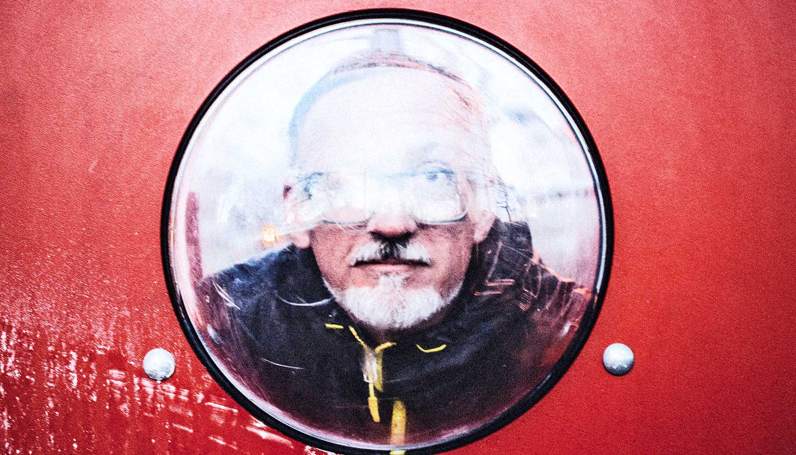
A new invention could give researchers a dynamic tool to study the brain’s role in various behaviors.
It’s a neuroscientist’s dream: being able to track the millions of interactions among brain cells in animals that move about freely, behaving as they would under natural circumstances. The new technology is a step towards realizing that goal.
Although designed for use with mice, information gleaned from the technology could someday shed light on neuronal activity in humans as well, says Alipasha Vaziri, who led the its development as head of the Laboratory of Neurotechnology and Biophysics at Rockefeller University. For example, it might allow us to better understand the neuronal basis of brain disorders such as autism and schizophrenia.
Vaziri says the tool provides an opening to an exciting range of discoveries. As an animal moves about its environment, for instance, some neurons direct spatial navigation while others receive sensory feedback from changes to the body’s position or the visual system.
“Until now, no one has been able to detect how these different neurons, which can be located at different depths within a volume of brain tissue, dynamically interact with each other in a freely moving rodent,” says Vaziri. Similarly, the tool can be used to record the interplay among neurons when two animals meet and interact socially.
Light show
The technology consists of a tiny microscope attached to a mouse’s head and outfitted with a specialized group of lenses called a microlens array.
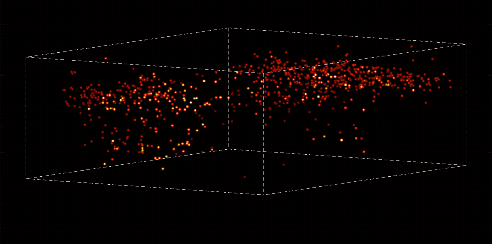
These lenses enable the microscope to capture images from multiple angles and depths on a sensor chip, producing a three-dimensional record of neurons blinking on and off as they communicate with each other through electrochemical impulses. (In the experiments, the mouse neurons are genetically modified to light up when they become activated.) A coaxial cable attached to the top of the microscope transmits the data for recording.
The head-mounted gear weighs only four grams, but Vaziri expects that planned modifications will make it even lighter.
Once the microlens array captures sensor images from within a volume of brain tissue, the next challenge is to process this raw data. Brain tissue is opaque, making it difficult to pinpoint the source of each neuronal light flash. Vaziri’s team solved this problem, which is the result of so-called scattering, by developing a new computer algorithm.
Scientists see neurons fire in brain of flying bat
“The algorithm utilizes the statistical properties of neurons’ distribution in space and in activity,” Vaziri explains, “while extracting additional information from the scattered emission light. This enables their activity to be simultaneously and faithfully recorded within a volume despite the highly scattering tissue properties.”
The result is a clear image that shows individual neurons flashing in sequence.
A better way
Vaziri’s lab has previously applied this algorithm, known by the acronym SID, in studies in which researchers secured the heads of the mice in fixed positions. Their latest research is the first to demonstrate that researchers can use these inventions together with a tiny microscope called the Miniscope, which a collaborating team at the University of California, Los Angeles developed, to measure neuronal activity volumetrically in unconstrained animals.
The technology, if widely adopted, could offer several advantages over two-photon microscopy, a broadly used neuroscience tool. For example, two-photon microscopy records neuronal activity within individual focal planes—thin, virtual “slices” of the sample—that then are combined to create a three-dimensional image.
Holographic brain device would edit our senses and memories
In contrast, Vaziri’s method immediately captures data in three dimensions over an entire volume of tissue, making it faster and more effective.
Vaziri plans to continue developing tools to record neuronal activity in even larger portions of the brain than currently possible, and at higher speeds and resolution.
“We hope this work will ultimately lead to a deeper understanding of how the brain processes information underlying the generation of behavior,” he says.
A paper describing the work appears in Nature Methods.
Source: Rockefeller University