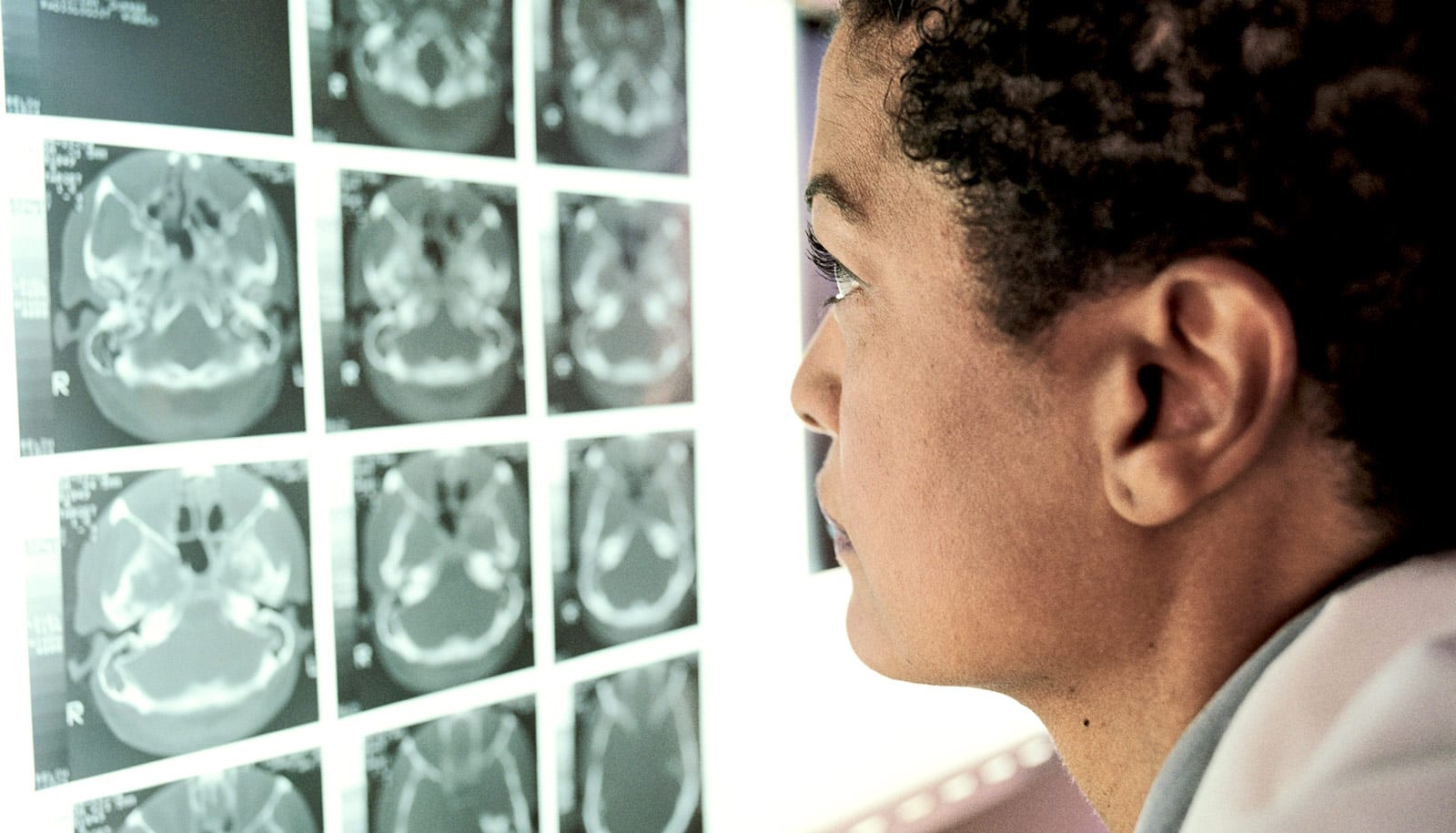
CT scans help identify organs at risk of injury from radiation therapy. A new automated technique uses a deep-learning algorithm to make the process easier.
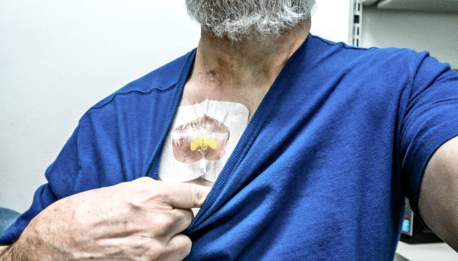
Radiation therapy is one of the most widely used cancer treatments, but a drawback of the procedure is that it can cause collateral damage to healthy tissue in proximity to cancerous growths.
The work appears in Nature Machine Intelligence.
“Using our model, it’s possible to delineate an entire scan in a few seconds, a task that would take a human expert over half an hour,” says coauthor Xiaohui Xie, professor of computer science at the University of California, Irvine.
“On a data set of 100 CT scans, our deep-learning method achieved an average similarity coefficient of more than 78%, a significant improvement over analyses done by radiation oncologists.”
The researchers focused on the head and neck for their study because of the complex anatomical structures and dense distribution of organs in this part of the body.
Also, unintended irradiation to sensitive tissue in this area can lead to adverse side effects such as difficulty in opening the mouth, deterioration of vision and hearing, and cognitive impairment.
Xie says the success of his team’s approach is attributable to the model’s two-stage design. First the system identifies regions containing vital organs, and then it extracts image features from these focal areas.
“Our deep-learning neural network greatly enhances the ability to delineate anatomies even with low-contrast CT scans,” Xie says. “And the setup is more computationally efficient than other methods, enabling it to be done with more standard levels of graphics processing unit memory. This means the technique can be deployed more readily in actual clinics.”
Xie’s collaborators are from China’s Shanghai Jiao Tong University School of Medicine and DeepVoxel Inc. of Costa Mesa.
Source: UC Irvine