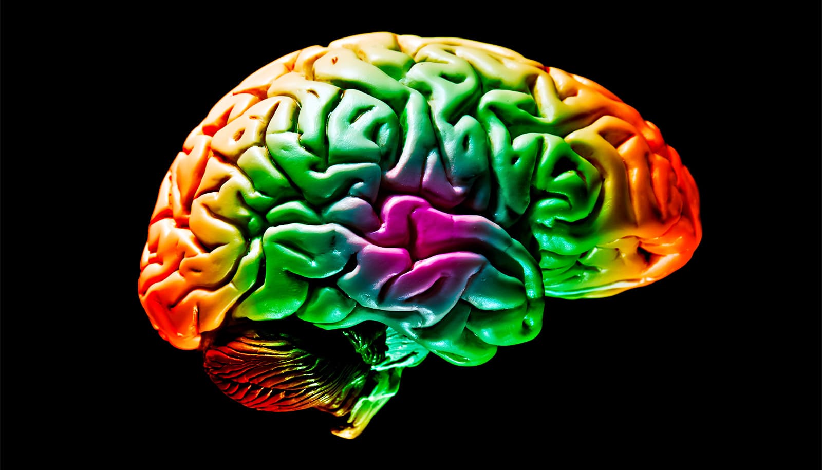
A new bioluminescence imaging technique has created highly detailed, and visually striking, images of the movement of oxygen in the brains of mice.
The human brain consumes vast amounts of energy, which is almost exclusively generated from a form of metabolism that requires oxygen. While the efficient and timely delivery of oxygen is known to be critical to healthy brain function, the precise mechanics of this process have largely remained hidden from scientists.
The new method, which can be easily replicated by other labs, will enable researchers to more precisely study forms of hypoxia in the brain, such as the denial of oxygen to the brain that occurs during a stroke or heart attack. The new research tool is already providing insight into why a sedentary lifestyle may increase risk for diseases like Alzheimer’s.
“This research demonstrates that we can monitor changes in oxygen concentration continuously and in a wide area of the brain,” says Maiken Nedergaard, codirector of the Center for Translational Neuromedicine (CTN), which is based at both the University of Rochester and the University of Copenhagen.
“This provides us a with a more detailed picture of what is occurring in the brain in real time, allowing us to identify previously undetected areas of temporary hypoxia, which reflect changes in blood flow that can trigger neurological deficits.”
The new method employs luminescent proteins, chemical cousins of the bioluminescent proteins found in fireflies. These proteins, which have been used in cancer research, employs a virus that delivers instructions to cells to produce a luminescent protein in the form of an enzyme. When the enzyme encounters a second chemical compound, a substrate called furimazine, the chemical reaction generates light.
Like many important scientific discoveries, employing this process to image oxygen in the brain was stumbled upon by accident. Felix Beinlich, an assistant professor in the CTN at the University of Copenhagen, had originally intended to use the luminescent protein to measure calcium activity in the brain. It became apparent that there was an error in the production of the proteins, causing a months-long delay in the research.
While Beinlich waited for a new batch from the manufacturer, he decided move forward with the experiments to test and optimize the monitoring systems. The virus was used to deliver enzyme-producing instructions to astrocytes, ubiquitous support cells in the brain that maintain the health and signaling functions of neurons, and the substrate was injected into the brain via a craniotomy. The recordings revealed activity, identified by a fluctuating intensity of bioluminescence, something that the researchers suspected, and would later confirm, reflected the presence and concentration of oxygen.
“The chemical reaction in this instance was oxygen dependent, so when there is the enzyme, the substrate, and oxygen, the system starts to glow,” says Beinlich.
While existing oxygen monitoring techniques provide information about a very small area of the brain, the researchers were able to observe, in real time, a large section of the cortex of the mice. The intensity of the bioluminescence corresponded with the concentration of oxygen, which the researchers demonstrated by changing the amount of oxygen in the air the animals were breathing. Changes in light intensity also corresponded with sensory processing. For example, when the mice’s whiskers were stimulated with a puff of air, the researchers could see the corresponding region of the brain light up.
The brain cannot survive long without oxygen, a concept demonstrated by the neurological damage that quickly follows a stroke or heart attack. But what happens when very small parts of the brain are denied oxygen for brief periods?
This question was not even being asked by researchers until the team in the Nedergaard lab began to look closely at the new recordings. While monitoring the mice, the researchers observed that specific tiny areas of the brain would go dark, sometimes for minutes, meaning that the oxygen supply was being cut off.
Oxygen is circulated throughout the brain via a vast network of arteries and smaller capillaries—or microvessels—which permeate brain tissue. Through a series of experiments, the researchers were able to determine that oxygen was being denied due to capillary stalling, which occurs when white blood cells temporarily block microvessels and prevent the passage of oxygen carrying red blood cells. These areas, which the researchers named “hypoxic pockets,” were more prevalent in the brains of mice during a resting state, compared to when the animals were active. Capillary stalling is believed to increase with age and has been observed in models of Alzheimer’s disease.
“The door is now open to study a range of diseases associated with hypoxia in the brain, including Alzheimer’s, vascular dementia, and long COVID, and how a sedentary lifestyle, aging, hypertension, and other factors contribute to these diseases,” says Nedergaard.
“It also provides a tool to test different drugs and types of exercise that improve vascular health and slow down the road to dementia.”
The research appears in the journal Science.
Additional authors are from the University of Rochester, the University of Copenhagen, and the University of Applied Sciences and Arts Northwestern Switzerland.
Support for the study came from the National Institute of Neurological Disorders and Stroke, the Dr. Miriam and Sheldon G. Adelson Medical Research Foundation, the Novo Nordisk Foundation, the Lundbeck Foundation, Independent Research Fund Denmark, and the US Army Research Office.
Source: University of Rochester