New research charts the radical evolutionary changes to the thigh bones of dinosaurs and birds that allowed them to stand on two feet.
The findings resolve a longstanding question about dinosaur evolution and offer a prime example of how new physical features can pop up briefly during embryonic development and then give way to older, known features in adults.

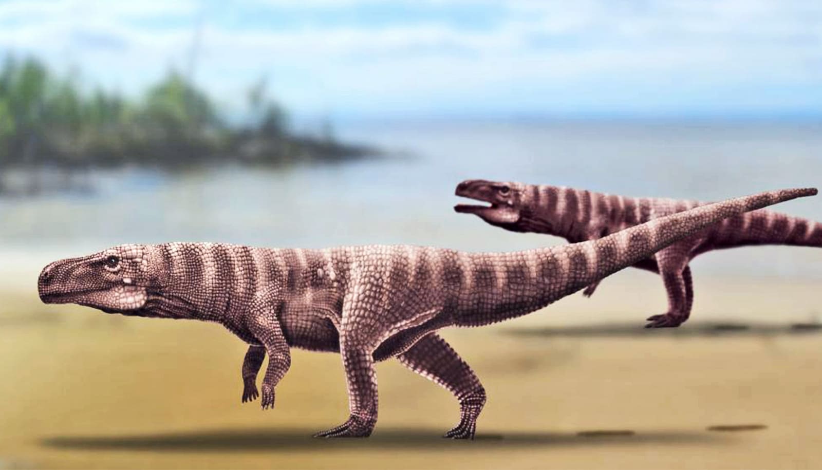
For the study, researchers focused on evolutionary shifts in the “femoral head”—where the upper femur connects to the hip—across a wide range of dinosaurs, early reptiles, and avians, and their modern-day counterparts.
The study appears in Proceedings of the Royal Society B.
“We figured out the way in which the femoral head, a critical part of dinosaur anatomy, developed,” says Bhart-Anjan S. Bhullar, associate professor of earth and planetary sciences in Yale University’s Faculty of Arts and Sciences and an associate curator at the Yale Peabody Museum of Natural History. “Inward-turned femoral heads are necessary for fast and effective, erect bipedal locomotion.”
For years, Bhullar says, there had been two conflicting theories about how dinosaur femoral heads developed. One theory held that the femoral head simply grew an attachment, or overhang, that reoriented the legs. The other theory was that the femoral head twisted inward over time.
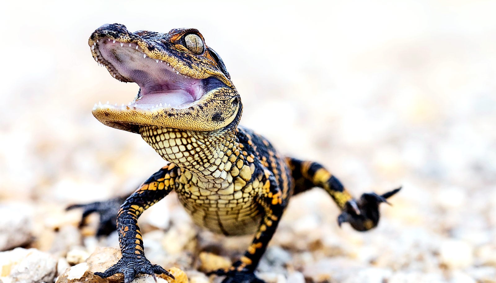
Evidence exists for both the twisting theory, found in early dinosaurs and modern crocodiles, and the in-growth theory, found in later dinosaurs and birds.
For the new study, the researchers used 3D imaging to study femoral head development in a variety of fossils and animal embryos. Bhullar’s lab at Yale is particularly known for its innovative use of CT scanning and microscopy to create 3D images of fossils.
What they discovered is that evidence for both theories occurs together.
“The embryonic development of this major feature changed completely—even while the feature itself remained constant in adults for quite some time,” Bhullar says.
“This sort of hidden shift in development might be more common than we think in evolution, and it should serve to caution against the widely held idea that features which develop differently must have evolved separately.”
Shiro Egawa, a former postdoctoral associate in Bhullar’s lab who is now a postdoctoral fellow at the National Center of Neurology and Psychiatry in Japan, is the study’s first author and co-corresponding author.
Additional coauthors are from the National Center of Neurology and Psychiatry in Japan, the American Museum of Natural History, Virginia Tech, the Royal Veterinary College in England, Pontificia Universidad Católica de Chile, Queensland Museum, CNRS/Muséum National d’Histoire Naturelle in France, Missouri State University, the RIKEN Center for Biosystems Dynamics Research in Japan, and Yale.
Funding for the research came, in part, from the National Science Foundation, the Japan Science Society, the Yamada Science Foundation, RIKEN, and the Zoological Society of Japan.
Source: Yale University