A new view of cholera’s “tail” could inform treatment.
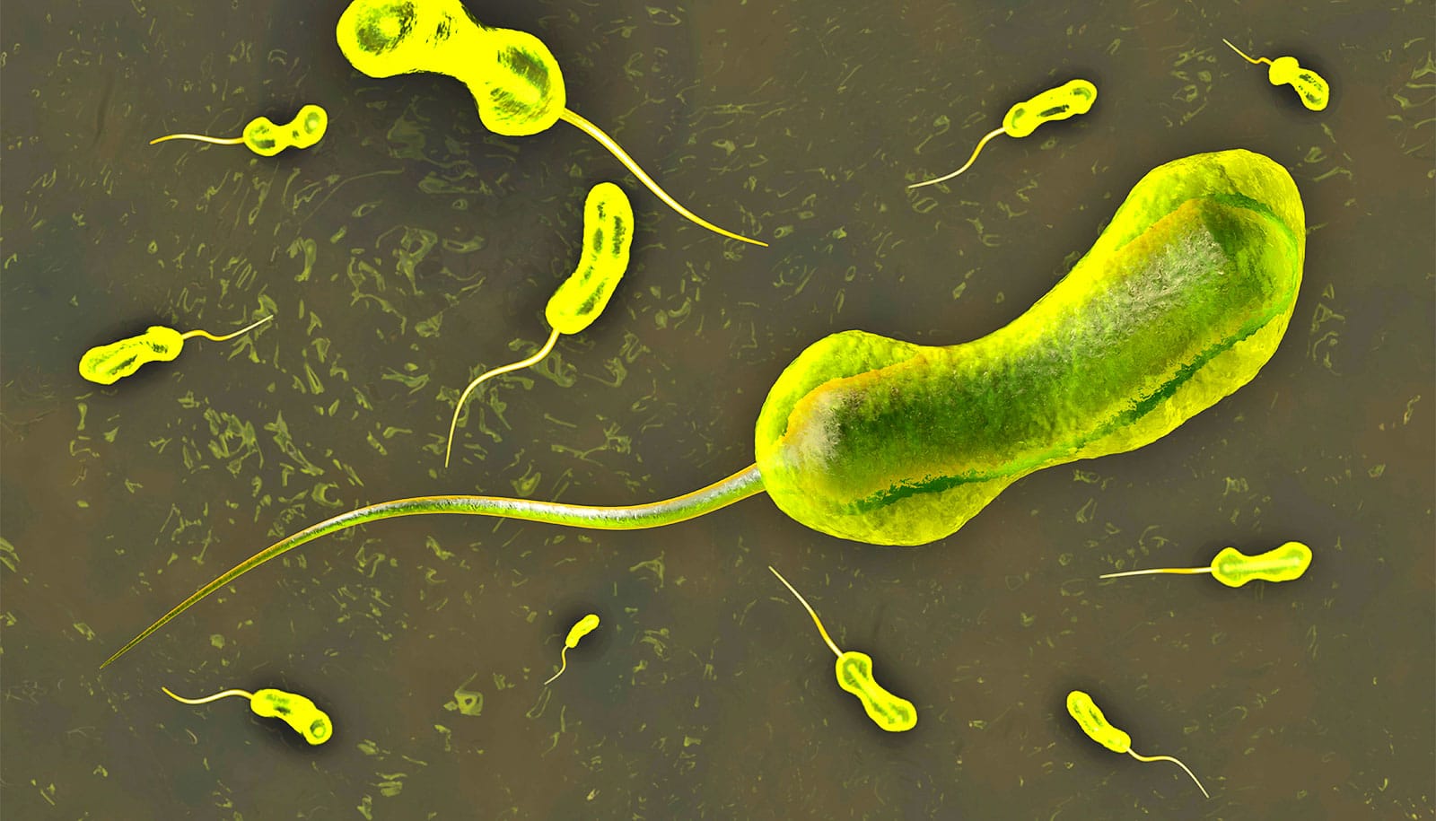
Cholera is a deadly bacterial disease that kills around 95,000 people every year. Vibrio cholerae bacteria infect cells in the small intestine, which the bacteria can do in part due to their flagella—powerful tail-like structures that the pathogen uses to move around.
Scientists have already identified the proteins and other molecules that make up V. cholerae‘s “tail.” But how these pieces fit together has remained unclear.
Now, new microscopy techniques have revealed the molecular structure of flagella in live V. cholerae bacteria, findings Yale School of Medicine (YSM) researchers reported recently in Nature Microbiology.
These findings could help researchers better understand how V. cholerae make their flagella and use it to get around, says Jun Liu, a professor of microbial pathogenesis at YSM and senior author on the study.
“To really understand the mechanism of the flagella—how they are able to assemble, how they rotate—you need near-atomic resolution,” says Liu, who is also a member of the Yale Microbial Sciences Institute at West Campus. These new results have allowed researchers to see cholera in “unprecedented detail,” he says.
Researchers have known about cholera’s unique flagella since the 1950s. Over the decades, scientists have found that these flagella—single thin structures that protrude out the ends of the bacteria—allow cholera to move unusually fast in liquid environments.
Scientists suspect that cholera’s speed and power help it push past the protective mucus layer in the small intestine to infect cells. The flagella are so important to infection that some cholera vaccines work by making the bacteria less mobile.
However, while researchers know what the flagella are made of, their underlying structure has so far eluded study. That’s in part because, unlike other bacteria, the four proteins that make up the flagella of V. cholerae are surrounded by a hydrophilic—or water loving—casing that prevents researchers from seeing the tail structure under the microscope.
There’s also the fact that current molecular techniques require researchers to kill bacteria and purify proteins to see what they’re made of. That means that researchers can see what the flagella are made of, but not how those components fit together.
As a result, the structure of V. cholerae‘s flagella is a “70-year mystery,” says Wangbiao Guo, PhD, a postdoctoral researcher in Liu’s lab and first author of the study.
One way to resolve the mystery is to get high-resolution images of flagella at a molecular level while V. cholerae is still alive. To do this, Guo and Sarah Zhang, a high school student working on the project over the summer, helped develop a new microscopy technique.
In collaboration with Merrill Asp—a postdoctoral researcher in the lab of Jing Yan, assistant professor of molecular, cellular, and developmental biology in Yale’s Faculty of Arts and Sciences—the researchers developed mutated V. cholerae whose flagella proteins were designed to light up. They then froze these bacteria in liquid ethane and used a powerful electron microscope to see the structure of the flagella at an almost atomic level.
These images allowed Liu and his colleagues to see how each of the four flagella proteins fit into specific spots inside the protective casing. The work revealed that the flagella of V. cholerae, while hidden behind the sheath, have a similar core structure but a different surface to the flagella of other bacteria. This suggests that the flagella of V. cholerae have evolved unique adaptations within the sheath.
So then, what makes cholera so much faster than other bacteria? One possibility is that the sheath itself might provide some lubrication. The structure of the flagellum suggests that it rotates independently from the outside casing. Since the sheath is hydrophilic, this might create a slippery system that helps the bacteria move through liquid more quickly than it otherwise would.
Figuring out how the flagella work will require more research, which can now be conducted thanks to these images and the techniques developed for this study, says Liu. The work could also set the stage for more drugs targeting cholera—either by going after the flagella, or by using these high-resolution images to look for other ways of attacking the disease, he says.
“We have at least provided some clues for the next development,” says Liu.
The research was supported by the National Institutes of Health, the National Science Foundation, and Yale University.
The content is solely the responsibility of the authors and does not necessarily represent the official views of the National Institutes of Health or National Science Foundation.
Additional support was provided by the Kleberg Foundation, Brown Foundation, Simons Foundation, Charles H. Revson Foundation, Damon Runyon Cancer Research Foundation, and San Antonio Area Foundation.
Source: Yale