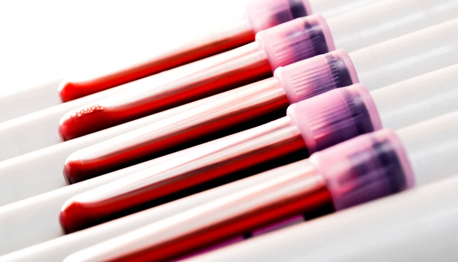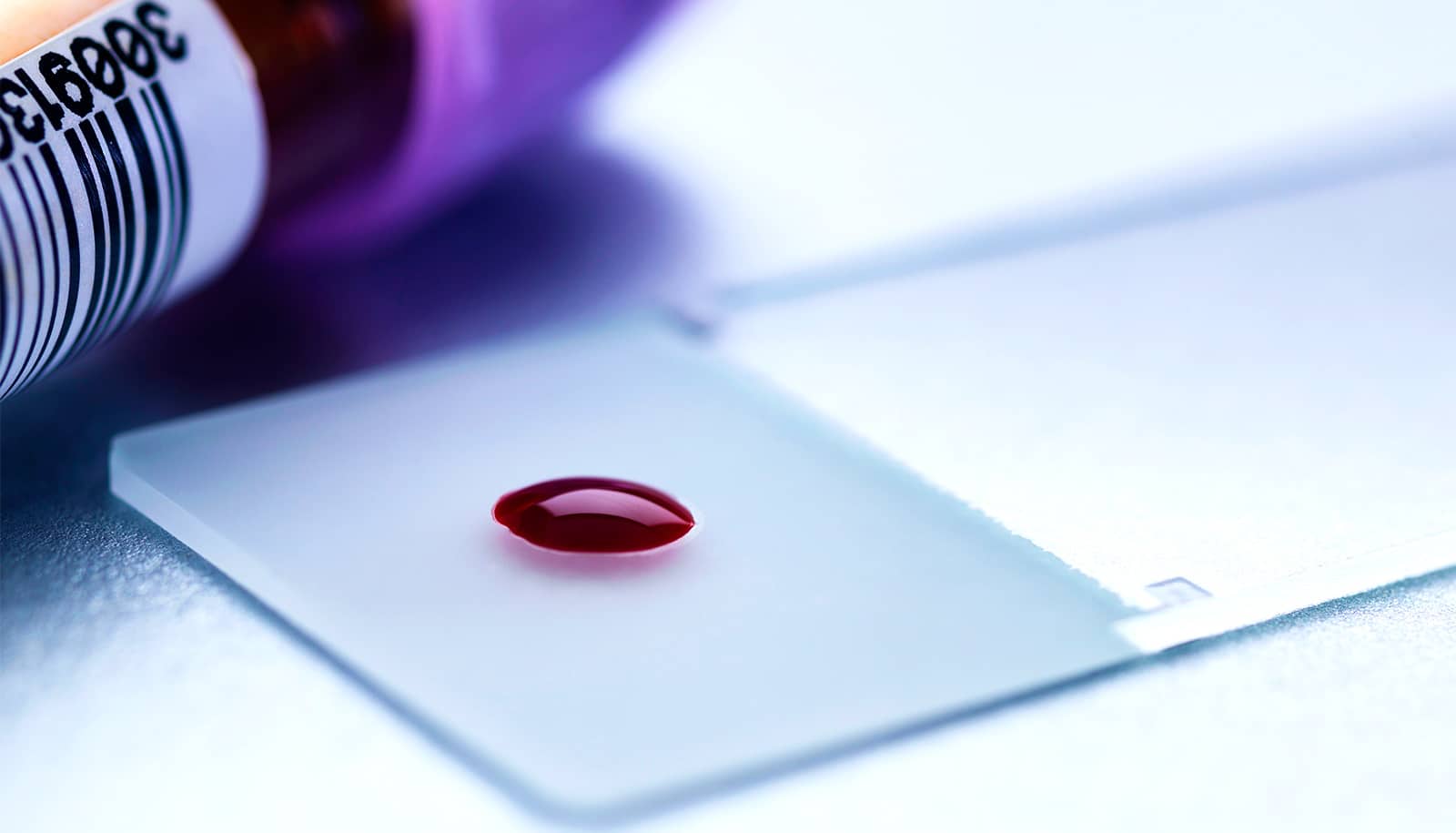
A new ultrasensitive diagnostic device could allow doctors to detect cancer quickly from a droplet of blood or plasma, report researchers.
The device could lead to timelier interventions and better outcomes for patients.
The “lab-on-a-chip” for liquid biopsy analysis detects exosomes—tiny parcels of biological information tumor cells produce to stimulate tumor growth or metastasize.
“Historically, people thought exosomes were like ‘trash bags’ that cells could use to dump unwanted cellular contents,” says lead author Yong Zeng, an associate professor of chemistry at the University of Kansas.
“But in the past decade, scientists realized they were quite useful for sending messages to recipient cells and communicating molecular information important in many biological functions. Basically, tumors send out exosomes packaging active molecules that mirror the biological features of the parental cells. While all cells produce exosomes, tumor cells are really active compared to normal cells.”
A million kitchen sinks
As reported in Nature Biomedical Engineering, the new device’s key innovation is a 3D nanoengineering method that mixes and senses biological elements based on a herringbone pattern commonly found in nature, pushing exosomes into contact with the chip’s sensing surface much more efficiently in a process called “mass transfer.”
“People have developed smart ideas to improve mass transfer in microscale channels, but when particles are moving closer to the sensor surface, they’re separated by a small gap of liquid that creates increasing hydrodynamic resistance,” Zeng says.
“Here, we developed a 3D nanoporous herringbone structure that can drain the liquid in that gap to bring the particles in hard contact with the surface where probes can recognize and capture them.”
“This is so simple and low-cost it has great potential to translate into clinical settings.”
Zeng compares the chip’s nanopores to a million little kitchen sinks: “If you have a sink filled with water and many balls floating on the surface, how do you get all the balls in contact with the bottom of the sink where sensors could analyze them? The easiest way is to drain the water.”
When scientists tested the chip’s design using clinical samples from ovarian cancer patients, they discovered the chip could detect the presence of cancer in a minuscule amount of plasma.
Easy and inexpensive
The microfluidic chips would also be cheaper and easier to make than comparable designs, allowing for wider and less-costly testing for patients.
“What we created here is a 3D nanopatterning method without the need for any fancy nanofabrication equipment—an undergraduate or even a high school student can do it in my lab,” Zeng says. “This is so simple and low-cost it has great potential to translate into clinical settings.”
Researchers could use the chip’s design to detect a host of other disease, Zeng says.
“Now, we’re looking at cell-culture models, animal models, and also clinical patient samples, so we are truly doing some translational research to move the device from the lab setting to more clinical applications.
“Almost all mammalian cells release exosomes, so the application is not just limited to ovarian cancer or any one type of cancer. We’re working with people to look at neurodegenerative diseases, breast, and colorectal cancers, for example.”
The National Institutes of Health and the University of Kansas Cancer Center’s Biospecimen Repository Core Facility, funded in part by a National Cancer Institute Cancer Center Support Grant, funded the work.
Source: University of Kansas