Burns cause approximately 180,000 deaths every year. A new method may offer a better way to predict the best treatment.
When a patient arrives at the emergency room with a burn, doctors do a clinical inspection to assess both the severity of the lesion in relation to the affected area of the body and the injury’s depth (first, second or third-degree burns).
The surgical team then makes a decision—based on this assessment and other considerations including bleeding and the extent of the damage—on how best to manage the patient’s care. However, because a wound can change in the days following the initial injury, using clinical inspection alone is inaccurate in 30 to 50 percent of cases.
Infrared thermography
Researchers suggest using non-invasive digital infrared thermography within the first three days after a patient suffers a burn could better predict the best mode of treatment. As reported in PLOS ONE, the non-invasive method is like taking a picture and is painless, quick, and requires minimal training.
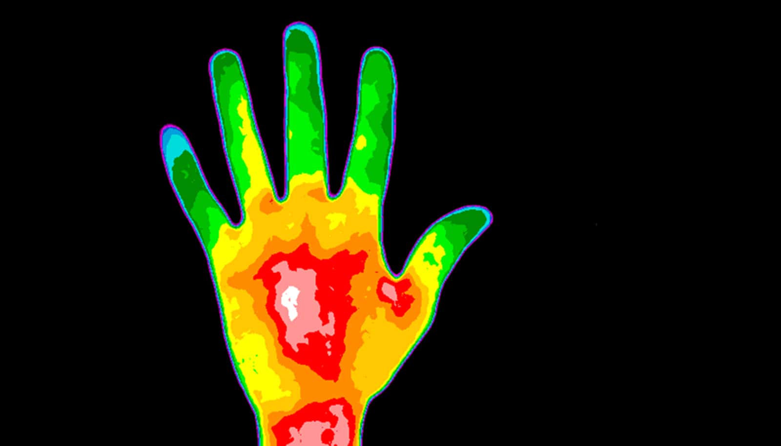
“Digital infrared thermography allows us to visualize the heat emitted by objects,” says corresponding author José Luis Ramírez García Luna, a PhD candidate in the experimental surgery department at McGill University.
“In the human body, heat emission depends on blood flow to the tissues, thus it is an indirect measurement of blood perfusion to the skin or a wound bed. Previous research has shown that the temperature of the skin correlates well with blood flow to it and that there are differences in the temperature pattern of healing vs. non-healing wounds.”
90% accuracy
The researchers devised a clinical prediction rule to decide which treatment a patient with a large burn in an extremity would receive based on the initial lesion’s thermal characteristics. To obtain the thermal characteristics of the lesion, they first obtained digital thermograms with a thermal camera and then recorded the difference in the average temperature of the burn and a patch of healthy adjacent skin.
After patient treatment and discharge, the researchers categorized treatment: re-epithelization if the wound healed by itself; skin graft if the patient received at least one; or amputation if extensive damage necessitated removal.
Finally, using machine learning, the researchers created a prediction model to categorize treatment based on the clinical data and the thermograms. They discovered the temperature difference between healthy skin and the wound alone is enough to make a prediction.
The algorithm predicted with 90 percent accuracy—both in the development cohort of patients, and as an independent validation cohort.
Lower costs
“We believe our method could become a useful tool for the early assessment of patients in the emergency department, as well as in the later stages of patient care to identify dead tissue, distinguish between partial and full-thickness burns, and detect complications such as the presence of infection or lack of blood flow,” says Ramírez García Luna.
“In our opinion, the most relevant contribution of our method to wound care is in allowing the clinical team to rapidly and objectively determine the treatment modality that is most likely needed, thereby preventing unnecessary procedures or delays in surgery.
“Because this has implications in selecting which patients may benefit more from certain interventions, and which will not, it is very likely that it can be translated to lowering costs related to treatment and hospital stay, as well as in reducing the length of hospital stay for the patients.”
Additional researchers are from McGill and Universidad Autonoma de San Luis Potosi.
Source: McGill University