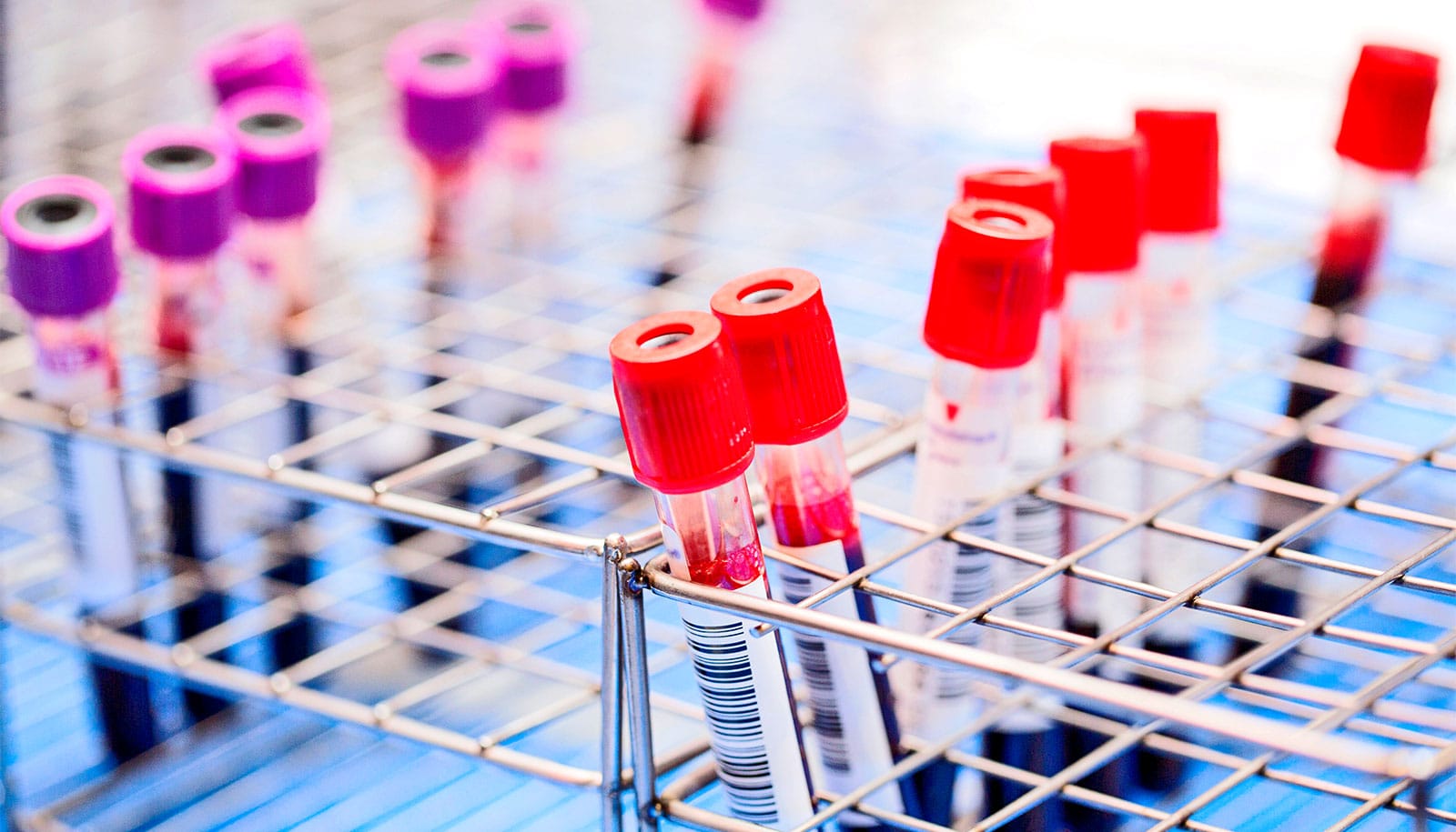
A new kind of microfluidic chip can capture rare, aggressive cancer cells, grow them on the chip, and release single cells on demand. The work could improve our understanding of how cancer spreads.
For the first time, researchers can easily compare two different “sister” cells—born of the same original cancer cell—to explore how different genes are activated and deactivated as cancer cells divide and spread. Studies with the new chip could also reveal why some cancer cells are resistant to drugs.
The ultimate goal of the project—led by Euisik Yoon, professor of electrical engineering and computer science at the University of Michigan—is to find out what drives the “self-renewal” processes that enable these aggressive cancer cells to behave like stem cells.
These cells are known as cancer stem cells. They are capable of dividing and turning into different kinds of cancer cells, with different genes turned on or off. Cancer researchers believe that if the stem-like properties can be switched off, the cancer will not be able to grow and spread.
“When a tumor forms, some cancer stem cells maintain stemness, while others are differentiated. By understanding this, we will know more about tumor formation and discover ways to inhibit it,” says Yu-Chih Chen, a research scientist in electrical engineering and computer science and co-first author of a paper in ACS Nano.
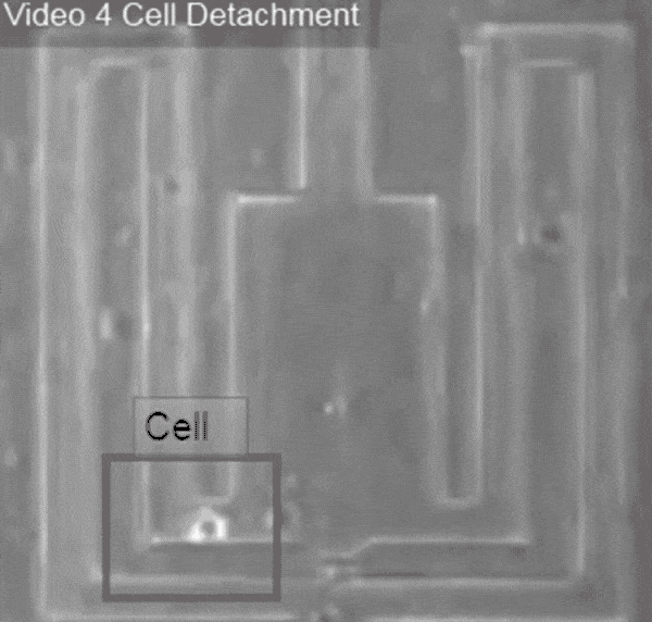
Pushed by a bubble
The base of the new chip is composed of carbon nanotubes covered in a plastic coating. When a cancer cell settles on the chip, it sticks itself to that coating. To release the cell, the researchers shone extremely short pulses of laser light near it. The light is readily absorbed by the carbon nanotubes, flash-heating them, while the plastic insulates the cell.
The heat causes trapped air between the nanotubes and plastic to expand, blowing a bubble under the cell. When the bubble bursts through the plastic, the cell detaches. Then, the cell can be flushed out of the chip and captured for genetic profiling.
Most existing methods for freeing individual captured cancer cells are either damaging to the cells or else cannot get them out of the chip reliably. The laser was precise enough that it could detach one side of a cell, leaving the other side anchored.
How glowing microRNA could illuminate cancer
And the bubble detachment process was so gentle that even surface proteins on the cell membrane were unscathed. The surface proteins are an important nondestructive avenue for identifying cancer stem cells.
Drug targets
To begin exploring the differences in gene expression between sister cells, the team first looked at a gene called Notch, which is associated with both normal and cancerous stem cells. If Notch was expressed in the daughter cells, it was a rough indication that the division was self-renewing. A Notch-positive cell could go on to produce two cells expressing the same gene, one Notch-positive and one Notch-negative, or two Notch-negative cells.
Their analyses demonstrated that Notch does not serve as a sole indicator for a cancer cell’s stem-like properties. Other genes associated with stem cells could be switched on or switched off in daughter cells with either Notch expression.
Algorithm learns to tell apart types of cancer cells
The task ahead of cancer researchers, with the help of the new chip, is to identify which of these genes are critical to a cancer stem cell’s self-renewing capabilities. If these can be shut down, forcing all cancer stem cells to produce only nonstem cells when they divide, it may be possible to subvert a tumor’s ability to grow and spread.
“If we identify some key genes, or a potential drug target, then pharmaceutical researchers can develop a compound to hit this drug target,” Chen says.
Drug testing inspired Yoon to develop this chip. On earlier chips, some cancer survived treatments, and he wanted to understand these cells better.
“Some cells are very resistant; some are easily killed,” says Yoon, who is also a professor of biomedical engineering. “We wanted to take individual cells out after drug screening and look at their genetic profiles to see if we can see what makes cancer cells stem-like.”
Future experiments could lead to what some cancer researchers call “functional cures,” similar to the management of HIV. The cancer doesn’t necessarily have to be eradicated. Stopping the cancer from spreading may be enough to enable a cancer patient to live a healthy life.
Partial funding for the work came from the Department of Defense and the National Institutes of Health.
Source: University of Michigan