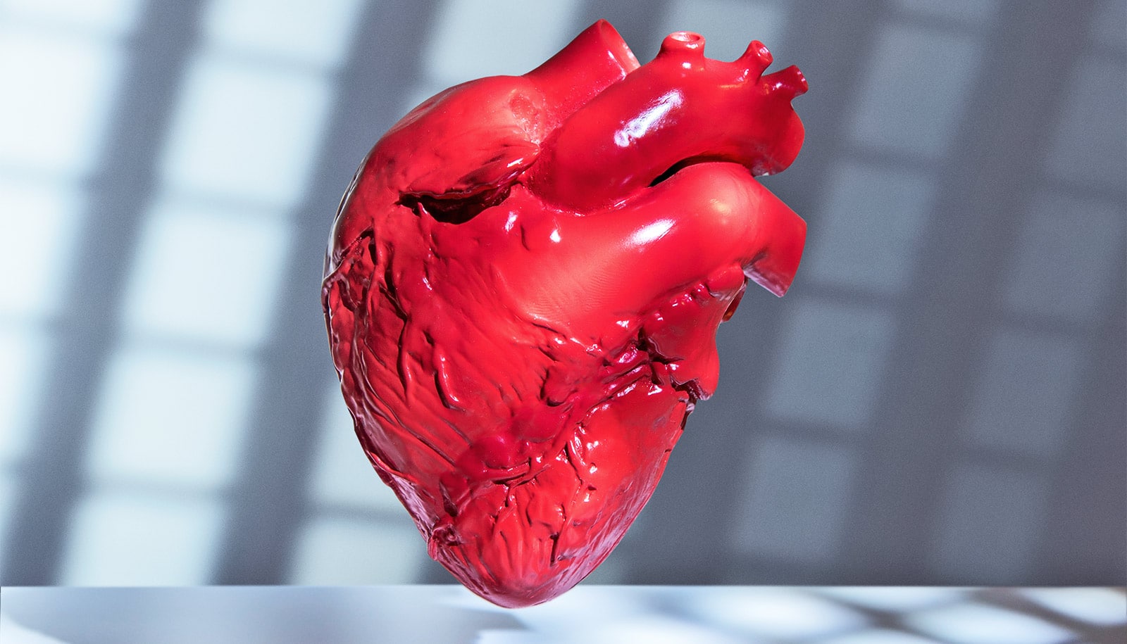
A new catheter probe combines two technologies to image the tiny arteries of a living heart.
To win the battle against heart disease, cardiologists need better ways to identify the composition of plaque most likely to rupture and cause a heart attack. Angiography allows them to examine blood vessels for constricted regions by injecting them with a contrast agent before X-raying them. But because plaque doesn’t always result in constricted vessels, angiography can miss dangerous buildups of plaque.
Intravascular ultrasound can penetrate the buildup to identify depth, but lacks the ability to identify some of the finer details about risk of plaque rupture.
This new catheter probe combines intravascular ultrasound (IVUS) with fluorescence lifetime imaging (FLIm) to image the tiny arteries of a living heart.
Professor Laura Marcu’s lab in the biomedical engineering department at the University of California, Davis, has now combined intravascular ultrasound with fluorescence lifetime imaging (FLIm). The new catheter can simultaneously retrieve structural and biochemical information about arterial plaque that could more reliably predict heart attacks.
The new device appears in a report in Scientific Reports.
This shows the artery in cross-section where the ultrasound (gray scale) depicts shape and structure and the fluorescence (colored band) depicts the composition of the artery. (Credit: UC Davis)
An optical fiber in the catheter sends short laser pulses into surrounding tissue, which fluoresces with tiny flashes of light in return. Different kinds of tissue (collagen, proteins, lipids) emit different amounts of fluorescence.
At the same time, an ultrasound probe in the catheter records structural information about the blood vessel.
The combination FLIm-IVUS imaging catheter provides a comprehensive insight into how atherosclerotic plaque forms, aiding diagnosis and providing a way to measure how plaques shrink in response to therapy.
The new catheter has been tested in living swine hearts and samples of human coronary arteries.
The catheter used in the study is flexible enough to access coronary arteries in a living human following standard procedures. It does not require any injected fluorescent tracers or any special modification of the catheterization procedures.
3D printing helps predict if new heart valves will leak
The new technique could not only can improve understanding of mechanisms behind plaque rupture—an event with fatal consequences—but also the diagnosis and treatment of patients with heart disease.
Marcu’s group is currently working to obtain FDA approval to test this new intravascular technology on human patients.
Source: Holly Ober for UC Davis