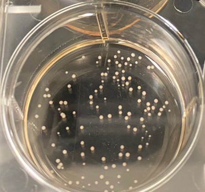
A new engineering feat expands what researchers can accomplish with organoids, including mini brains—the lab-grown balls of human cells that mimic some of a brain’s structure and functionality.
It could be the world’s tiniest EEG electrode cap, created to measure activity in a brain model the size of a pen dot. Its designers expect the device to lead to better understanding of neural disorders and how potentially dangerous chemicals affect the brain.
“This provides an important tool to understand the development and workings of the human brain,” says David Gracias, a chemical and biomolecular engineer at Johns Hopkins University. “Creating micro-instrumentation for mini-organs is a challenge, but this invention is fundamental to new research.”
Since organoids were first created more than a decade ago, researchers have modified stem cells to create small-scale kidneys, lungs, livers, and brains. Researchers use the complex, miniature models to study how the organs develop.
Humans “are not ‘Flat Stanley.’ Flat measurements have inherent limitations.”
Researchers study unaltered organoids next to ones that are genetically modified, injected with viruses, and exposed to chemicals. Organoids, particularly mini brains, are increasingly important in medical research because they can be used in experiments that would otherwise require human or animal testing.
But because the conventional apparatus to test organoids is flat, researchers have been able to examine only limited cells on their surface. Knowing what’s happening to a larger number of cells in the organoid would help reveal how organs function and diseases progress, Gracias says.
“We want to get information from as many cells as possible in the brain, so we know the state of the cells, how they communicate, and their spatiotemporal electrical patterns,” he says.
Humans “are not ‘Flat Stanley,'” says coauthor Lena Smirnova, a research associate in the environmental health and engineering department. “Flat measurements have inherent limitations.”
Inspired by the electrode-dotted skull caps used to detect brain tumors, the team created tiny EEG caps for brain organoids from self-folding polymer leaflets with conductive polymer-coated metal electrodes. The microcaps wrap around the entirety of an organoid’s spherical shape, enabling 3D recording from the entire surface so that, among other things, researchers can listen to the spontaneous electrical communication of neurons during drug tests.
The data is expected to be superior to the current readings from conventional electrodes on a flat plate.
“If you record from a flat plane, you only get recordings from the bottom of a 3D organoid sphere. However, the organoid is not just a homogeneous sphere,” says first author Qi Huang, a PhD candidate in the chemical and biomolecular engineering department. “There are neuron cells that communicate with each other. That’s why we need a spatial-temporal mapping of it.”
With more detailed information from organoids, researchers can study whether chemicals used in consumer products cause problems in brain development, says coauthor Thomas Hartung, director of the Center for Alternatives to Animal Testing at the Johns Hopkins Bloomberg School of Public Health.
“Some chemicals like pesticides are especially suspicious, as many kill insects by damaging their nervous system,” Hartung says. “Flame retardants are another class of chemicals where we have concerns.”
Researchers hope that readings from the caps could reduce the number of animals needed to test chemical effects. Traditional testing of just one chemical requires about 1,000 rats and costs about $1 million, Hartung says. The results from organoids are also more germane, he adds, because human brains are very different from rat and mice brains.
The study is published in Science Advances. Additional coauthors are from Syracuse University, the University of Washington, and Johns Hopkins.
The US Environmental Protection Agency, Johns Hopkins University Discovery Award, the National Institutes of Health/ National Institute of Biomedical Imaging and Bioengineering, and the National Institute on Drug Abuse supported the work.
Source: Johns Hopkins University