Scientists have created a simpler and faster method for focusing visible light through a range of materials that mirror human skin’s light-scattering properties. The work is a step toward using visible light to image inside the body.
Dense structures like bone show up clearly in X-rays, but softer tissues like organs and tumors are difficult to make out. That’s because X-rays are strongly deflected by bones, while they cut straight through soft tissue.
Visible light, on the other hand, is deflected by soft tissue. Until recently, this has made seeing through skin with visible light a nonstarter—while light can get through, it’s scattered every which way. At the same time, visible light would be safer for diagnostic imaging than higher-energy X-rays.

“Light comes in, it hits a molecule, hits another, hits another, does something really crazy, and exits this way,” says Moussa N’Gom, assistant research scientist in electrical engineering and computer science and first author on a study in Scientific Reports that explains the challenge of predicting the paths of individual light rays.
By understanding exactly how a patch of skin scatters the light, researchers hope to carefully pattern light beams so that they focus inside the body—a first step toward seeing into it.
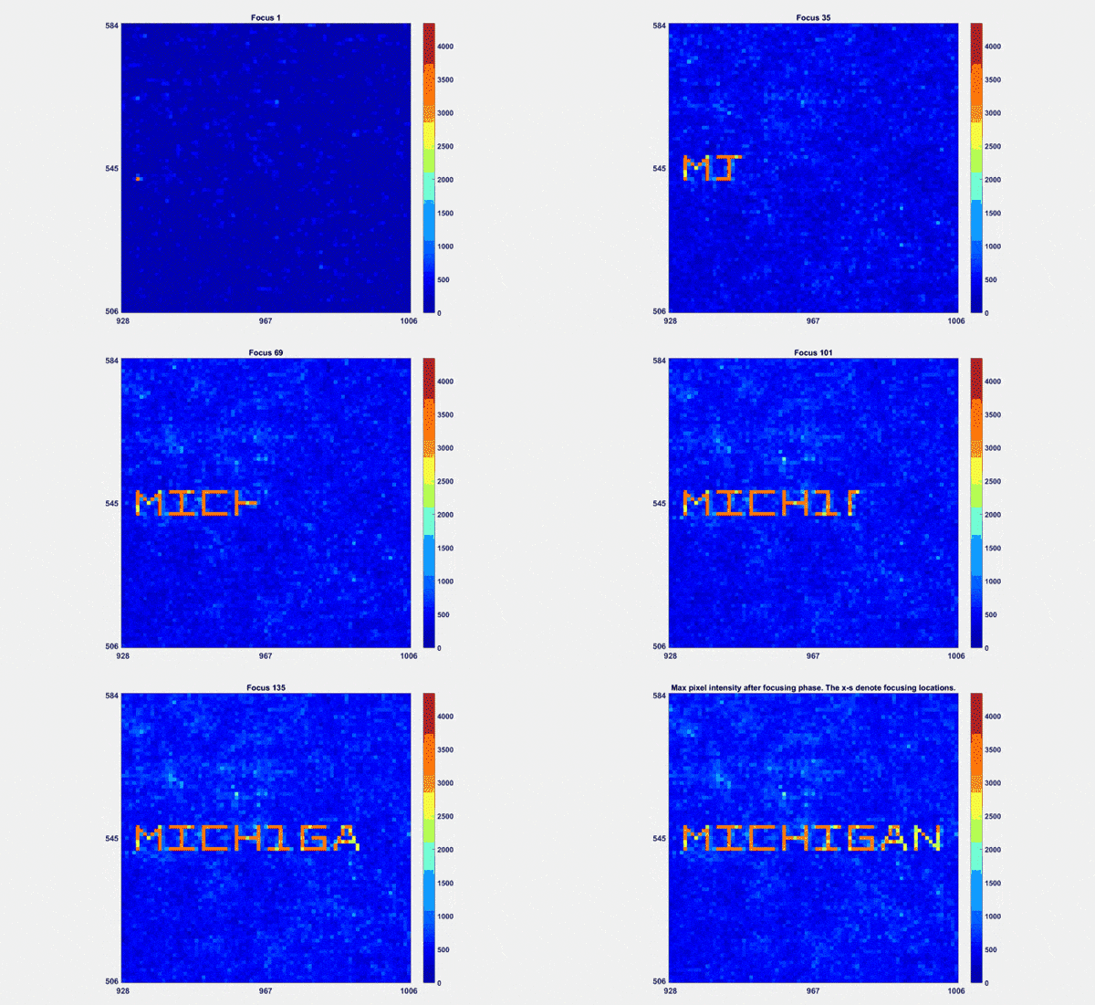
In their experiments, the researchers spelled “MICHIGAN” with a beam of light shone through yogurt and crushed glass. They chose those materials because they scatter light strongly and serve as good models for skin. Their demonstration, reminiscent of writing a name with a flashlight, shows that they can take a single, quick scan of the material and focus through it at many points—as they would need to do if imaging tissue inside the body.
Light and mirrors
The field of imaging objects through materials, from layers of paint to eggshells and even mouse skulls, has made great strides in the last decade. The typical “holographic” method untangles the scattering pattern by looking at how the light waves interfere with each other—this gives information about how different rays were delayed on their way through the material.
This method is very precise, says N’Gom, but it is slow. To speed things up, researchers typically figure out just enough of the scattering pattern to focus on a particular point. To focus on a different point, the material has to be scanned again. This would slow the process of measuring the size or texture of a tumor, for example.
“Our method is significantly faster and more convenient because we use a single set of measurements to generate all these points, and we don’t have to rescan,” N’Gom says.
Holographic scans of blood find malaria in cells
As is typical for focusing-through-materials experiments, the researchers used a spatial light modulator to produce patterns of light. If you shone a laser through frosted glass, it would enter at a point on one side, at a particular angle, and then leave the other side through many points, in different directions.
By combining a screen with an array of mirrors, a spatial light modulator can do the reverse, sending light to a surface at many points, at many angles, so that these rays converge on a point on the other side of the material.
They set up the spatial light modulator to shine in hundreds of different patterns (461 in all). But rather than analyzing the paths of individual light rays emerging from the other side, N’Gom’s team measured the brightness—how much light made it out.
They developed an algorithm to trawl through the incoming light patterns and outgoing brightness measurements, using the information to build up a mathematical representation of the material’s scattering pattern, called the transmission matrix.
“Previous techniques, instead, used complex so-called holographic setups to extract the necessary information,” says Raj Rao Nadakuditi, associate professor of electrical engineering and computer science and senior author on the study. “We were able to achieve the same through simple brightness measurements and as a result operate much faster.”
Using the transmission matrix, N’Gom’s team could figure out exactly how to set the spatial light modulator to get a bright spot at any point on the other side of the ground glass or yogurt.
In the yogurt, there was a time limit on how long the map was good—just a few minutes. It was enough time for N’Gom and his colleagues to spell “MICHIGAN” in 157 shots.

Ready in 5 years?
In skin, the time constraints are much tighter—they would need a new map about every millisecond. Even so, with state-of-the-art electronics, N’Gom thinks their algorithm could run that fast.
Laser scalpel would leave skin unharmed
Another challenge in seeing through skin is that they wouldn’t be able to position a detector beneath it to measure the brightness of the light. For this, N’Gom says that researchers are using ultrasound to detect heating in the target tissue—a measure of how much light is getting through.
Finally, with the light focused inside, an imaging device would still need to focus the light coming back out of the skin. For this, they could essentially run the pattern of light back through the transmission matrix to deduce where the reflection was coming from.
Considering recent progress and ongoing studies in focusing light through translucent materials, N’Gom anticipates that we may see the first visible light images taken through skin within the next five years.
The Defense Advanced Research Projects Agency supported the study work.
Source: University of Michigan


